Metastatic Melanoma
Findings:
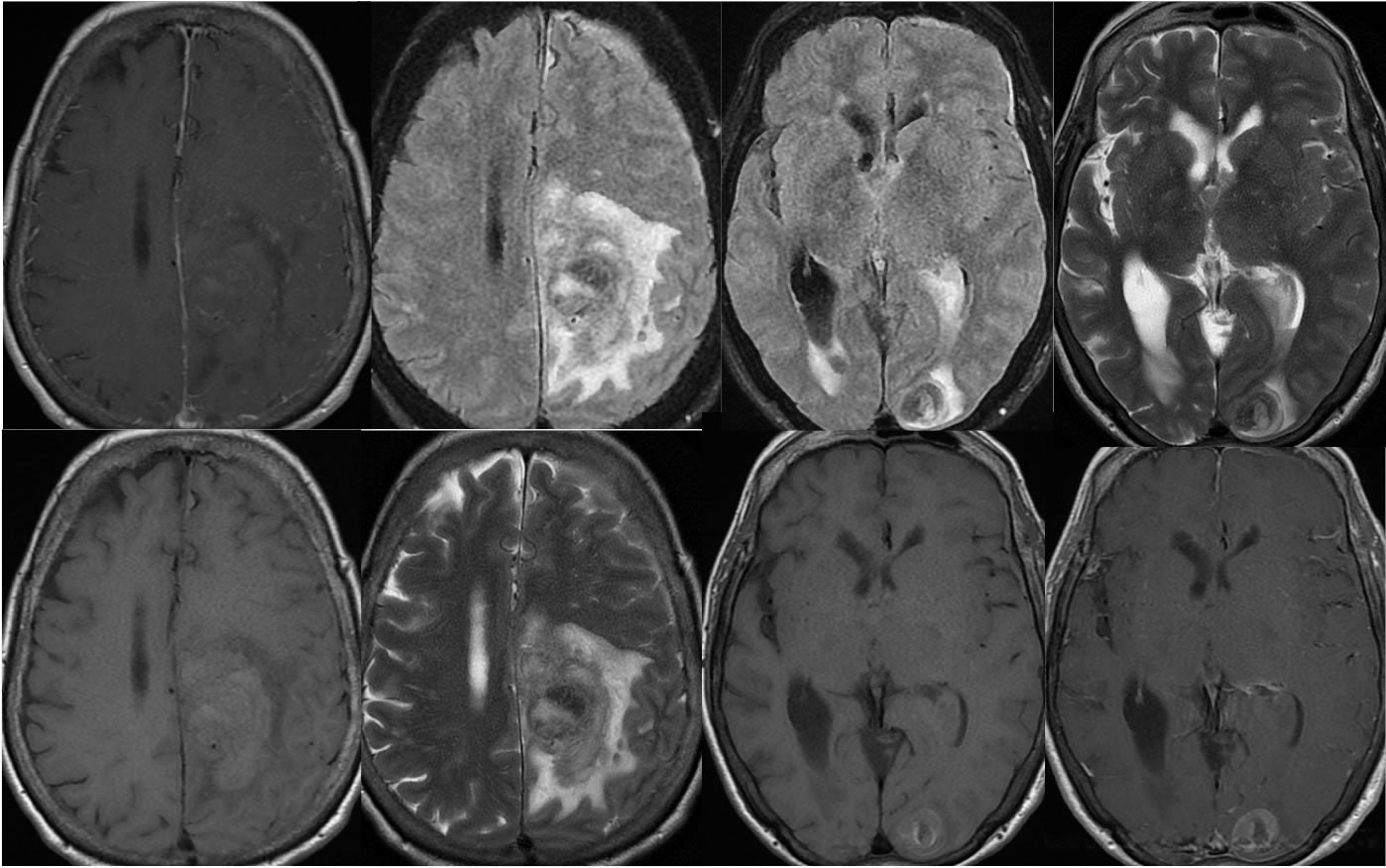
Multiple MR images demonstrate a large parenchymal hematoma in the left posterior frontal parasagittal region. An additional lesion with hemorrhagic change and nodular enhancement involves the left occipital lobe. Associated intraventricular hemorrhage is present. While no definite nodular enhancement is associated with the dominant parenchymal hematoma, there is a zone of T1 precontrast hyperintensity which demonstrates increased T2 signal. The left occipital lesion is also associated with T1 precontrast hyperintensity.
Differential Diagnosis/Discussion:
When intracranial hemorrhage is found, a workup is undertaken for underlying cause. The presence of nodular enhancement within a separate lesion indicates the presence of metastatic disease. T1 precontrast hyperintensity can sometimes represent subacute hemorrhagic products, but in the presence of metastatic disease, melanin causing T1 shortening indicates that melanoma is the underlying primary tumor.
BACK TO
MAIN PAGE