Metastatic Goblet Cell Carcinoid
Findings:
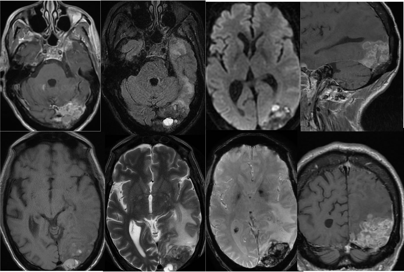
Multiple MR images demonstrate a poorly defined irregular enhancing mass with hemorrhagic changes in the left occipital lobe which is inseparable from but does not include the left transverse sinus. Surrounding vasogenic edema and mass effect is present. This lesion has a broad base of contact to the dura and is difficult to determine whether intaaxial or extraaxial. A smaller peripheral mass of similar characteristics projects over the left anterior temporal lobe.
Differential Diagnosis:
The presence of multiple lesions raises the high likelihood of metastatic disease. If the left occipital mass were solitary, the differential diagnosis still would primarily include metastatic disease, that other high-grade malignancy such as a meningeal sarcoma, malignant meningioma, or glioblastoma with dural invasion could be considered.
Discussion:
Goblet cell carcinoid is a rare and aggressive GI tract malignancy found in the appendix, which histologically has biphasic population of malignant goblet cells with adenocarcinoma differentiation and malignant neuroendocrine cells. No specific features of the metastases above allow a prospective concern for goblet cell carcinoid.
BACK TO
MAIN PAGE