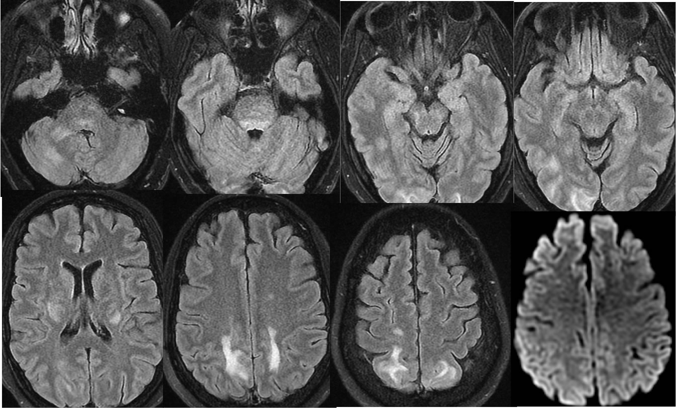
PRES
Findings:
Multiple MRI FLAIR and diffusion weighted images demonstrate patchy signal abnormalities in the bilateral parietooccipital subcortical regions which are nearly symmetric. Additional signal abnormalities are present in the bilateral basal ganglia, dorsal pons, and right cerebellar white matter. No restricted diffusion is seen.
Differential Diagnosis:
The symmetric bilateral parietal subcortical involvement should raise the possibility of PRES. Additional distribution discussed above is not atypical for PRES. Other causes such as encephalitis or multifocal infarction could appear similar, but there is no restricted diffusion. Clinical history is often helpful for distinction.
Discussion:
-Other example of PRES with discussion
BACK TO
MAIN PAGE