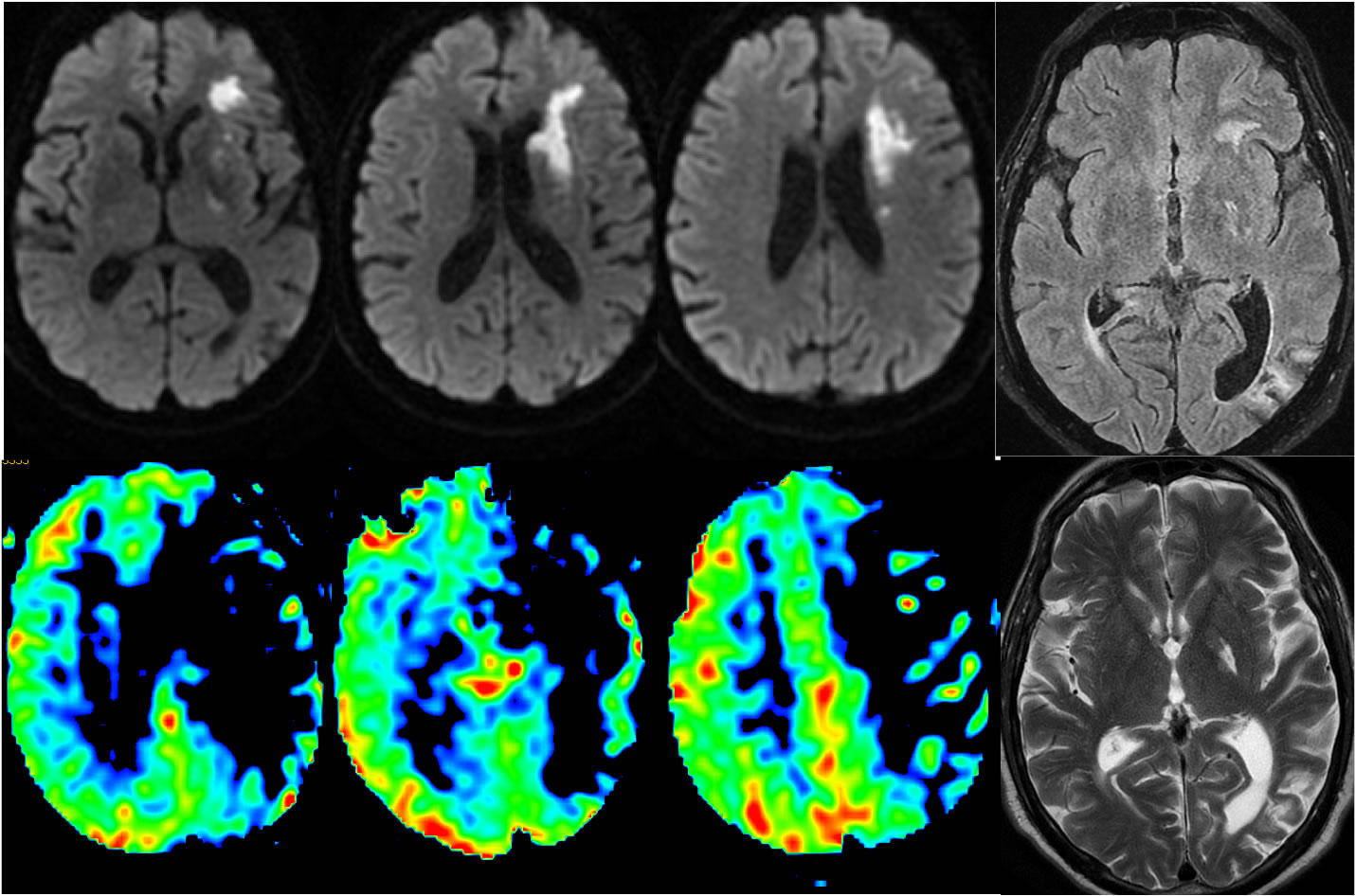
L MCA Infarct with Penumbra
Findings:
Patchy zones of restricted diffusion are present within the deep left frontal white matter. T2 and FLAIR images demonstrate superimposed remote infarcts in the left posterior limb internal capsule and left temporo-occipital region. Arterial spin labeling demonstrates a broad zone of hypoperfusion throughout the left MCA territory that is much larger than the zone of acute infarction.
Discussion:
This is a good example of a combination of acute infarction, remote infarction, and penumbra at risk territory. Arterial spin labeling when available is a rapid and noninvasive technique to assess perfusion on MR.
BACK TO
MAIN PAGE