Reducible Anterior TMJ Disc Displacement
Findings:
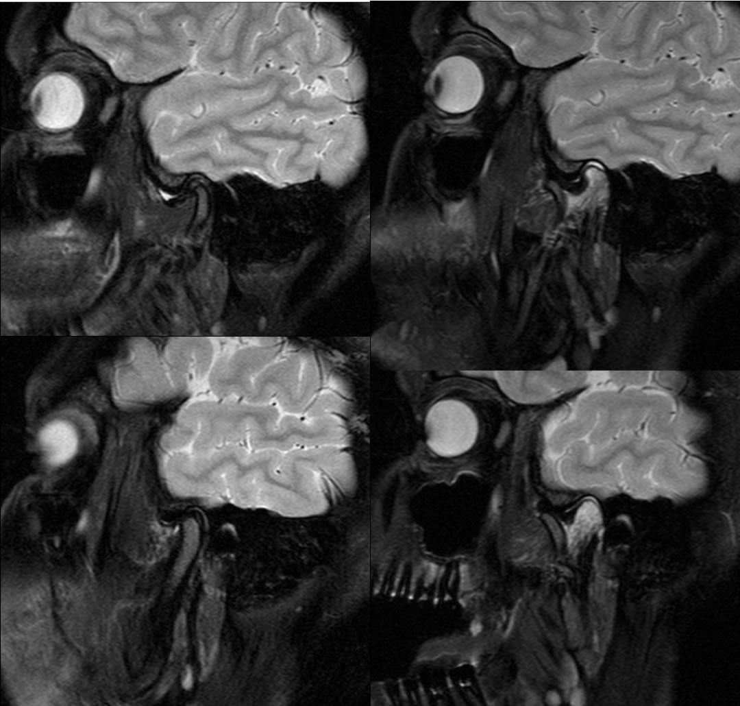
Oblique sagittal T2 fat saturated images demonstrate anterior displacement of the left TMJ articular disc in the closed mouth position, which recaptures in the open mouth position with normal range of motion of both TM joints. There is normal signal intensity and morphology of the right TMJ articular disc.
Discussion/Differential Diagnosis:
The normal TMJ articular disc demonstrates low signal intensity on T2 images and has a biconcave meniscoid shape. With closed mouth positioning, the disc should have its posterior band near the 12 o'clock position with relation to the mandibular condyle. With jaw opening, the disc rotates anteriorly and the condyle glides over the undersurface to rest near the 6 o'clock position of the condylar eminence. Nonreducible disc displacements may result in pain and limited range of motion, while reducible disc displacements may cause a clicking sensaton which may or may not be associated with pain.
BACK TO
MAIN PAGE