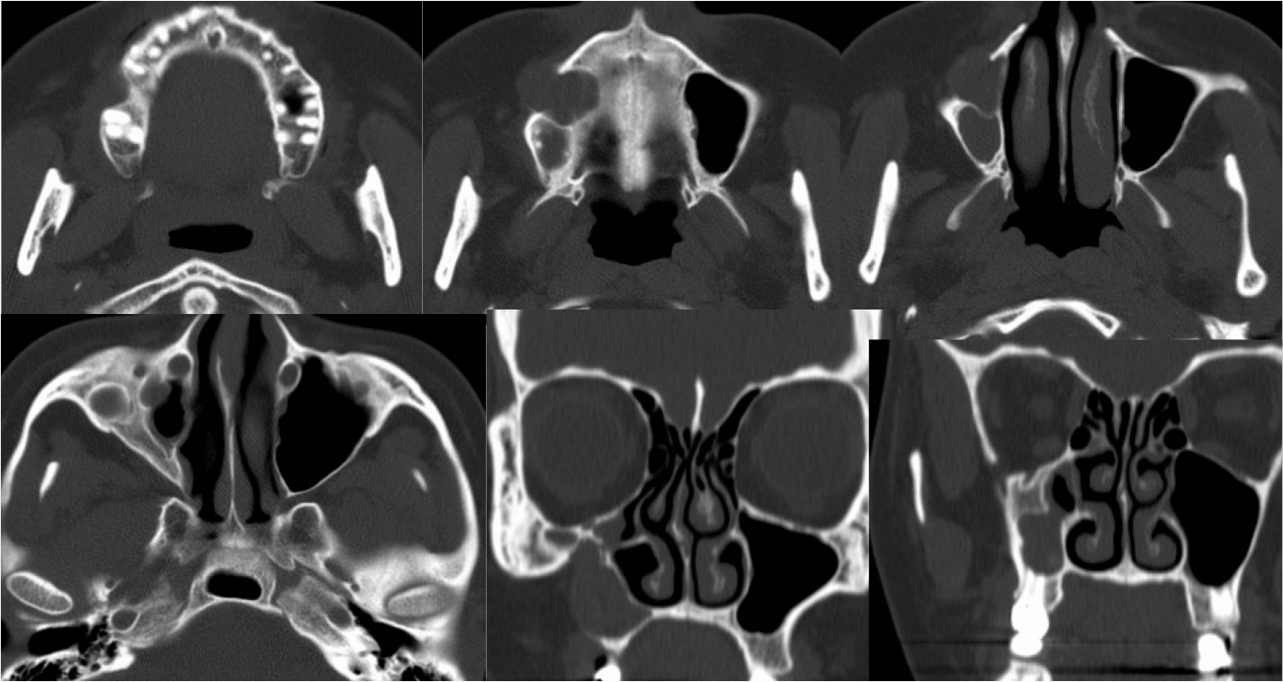
Chronic R maxillary sinusitis with mucocele
Findings:
Axial and coronal CT images demonstrate asymmetric decreased volume of the right maxillary sinus with wall thickening and overall moderate opacification. More anteriorly in the right maxillary sinus, there is an expansile low density lesion with bone interruption laterally and associated bone remodeling. No definite periapical lucencies are seen in association with the maxillary teeth.
Discussion/Differential Diagnosis:
The differential diagnosis for an expansile lesion of the paranasal sinuses is broad including benign and malignant etiologies, but may be narrowed by the presence of smooth bone remodeling which would indicate a long standing likely benign process. Mucocele results from an obstructed sinus cellule that fills and expands with secretions. Bone interruptions may occur when the expansion outpaces the periosteal repair process and this may simulate a more aggressive process. Mucoceles are most common in the frontal and ethmoid sinuses, less common in maxillary and sphenoid sinuses. THe presence of a sphenoid sinus mucocele should raise suspicion for an obstructive mass at the sphenoethmoid recess. Contents of the mucocele may have variable CT attenuation and MR signal due to the protein concentration and presence of blood products, but nodular enhancement is not seen.
BACK TO
MAIN PAGE