Left Mesial Temporal Sclerosis
Findings:
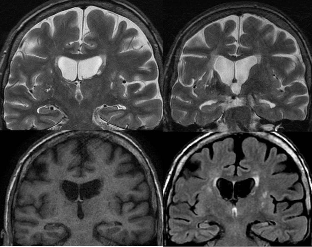
Coronal thin section MR images through the temporal lobes, including axial T2, axial T1, and FLAIR demonstrate decrease volume of the left hippocampal formation, associated with FLAIR hyperintensity and loss of internal architecture.
Discussion/Differential Diagnosis:
Hippocampal volume loss and gliosis may occur from remote insult such as infarct or previous inflammatory process, or be idiopathic. This case is not expansile, therefore unlikely to represent neoplasm or active limbic or other encephalitis. The association with chilhood febrile seizures is controversial. Mesial temporal sclerosis is the most common association with intractible temporal lobe epilepsy. Up to 10% may be bilateral. In addition to the volume loss and gliosis, there is loss of internal architecture with nonvisualization of the stratum radiata. Other portions of the limbic system may show ipsilateral volume loss, including but not limited to the fornical columns and mamillary bodies. Multiple other structures may show volume loss. If medical management is ineffective, anterior temporal lobectomy may be successful in up to 90% of patients.
BACK TO
MAIN PAGE