Hemangiopericytoma
Findings:
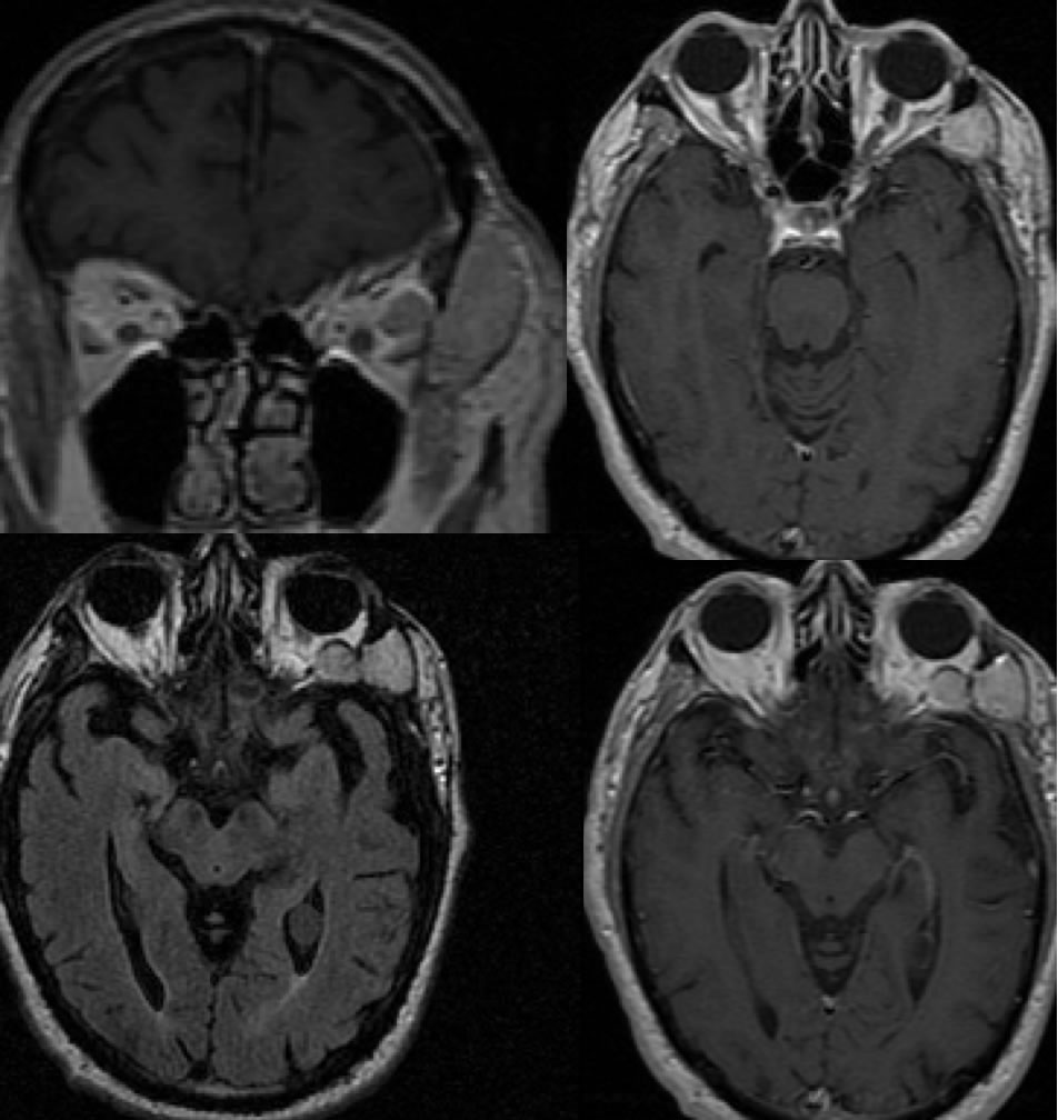
Axial FLAIR and axial/coronal T1 post contrast images demonstrate a bilobed homogeneously enhancing mass within the extraconal space of the left postseptal orbit, associated with a larger laterally projecting component extending through the lateral orbital wall displacing the temporalis muscle.
Discussion/Differential Diagnosis:
The differential diagnosis for an enhancing mass involving the orbit generally includes hemangioma, lymphoma, and metastasis. Nothing about this mass is specific for the diagnosis of hemangiopericytoma, but the extent through the lateral orbital wall would be unusual for lymphoma and would not be seen with a cavernous hemangioma. Orbital hemangiopericytomas (HPC) are uncommon and less than 3% of primary orbital tumors. This a highly vascular mass that has origin from pericytes, which grows slowly but does have malignant potential. There is some overlap in histology between HPC and solitary fibrous tumors (SFT). HPC can be considered a more agressive form of SFT. When involving the meninges, these are essentially indistinguishable from meningiomas and are treated the same way with resection with or without radiotherapy. HPC typically destroy bone while meningiomas more often cause hyperostosis.
BACK TO
MAIN PAGE