Squamous Cell Carcinoma, Tongue Base with Bilateral Nodal Metastases
Findings:
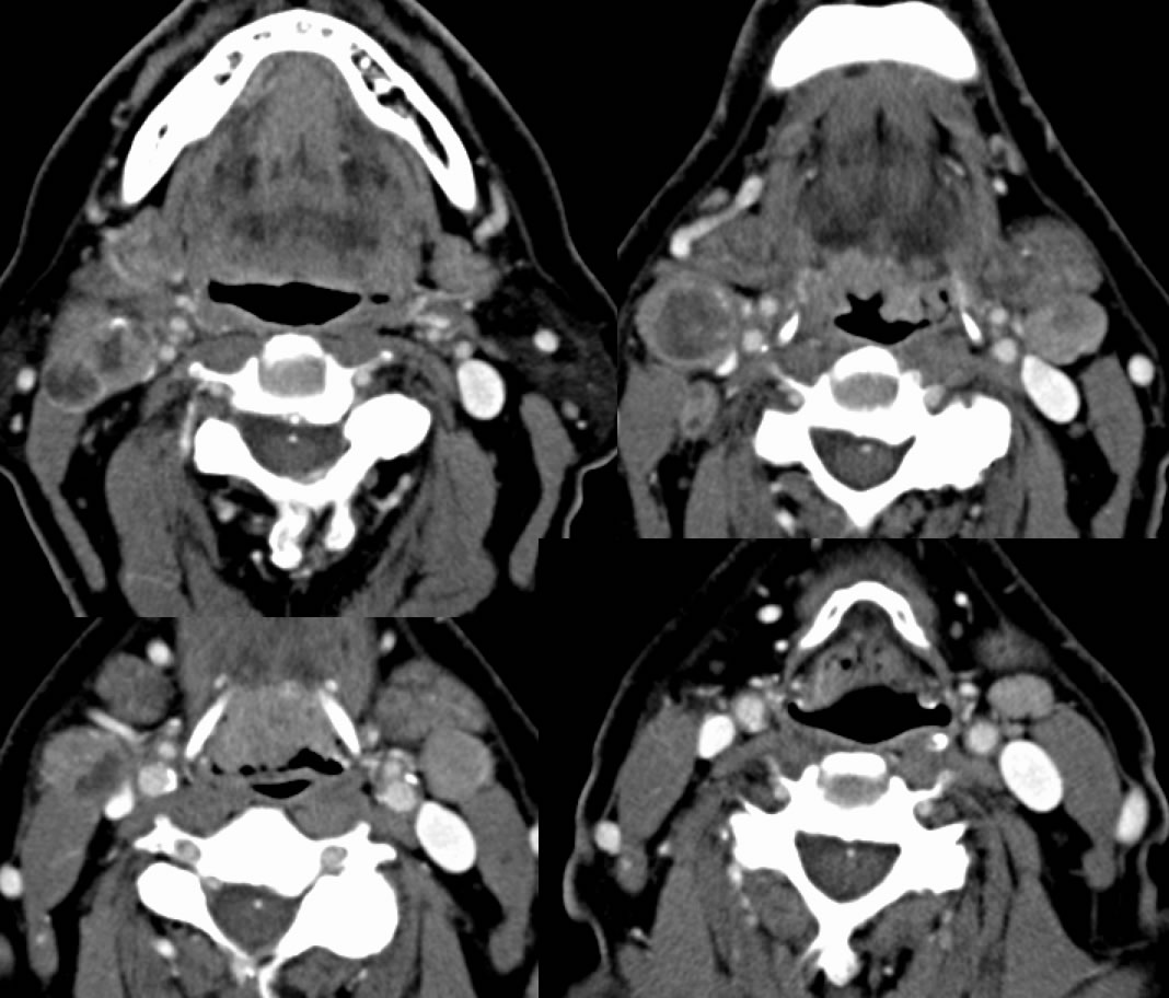
Axial contrast enhanced neck CT images demonstrate multiple enlarged solid and centrally necrotic lymph nodes in the bilateral level 2 chains as well as left level 3. Soft tissue mass with central ulceration involves the midline tongue base extending slightly to the right.
Discussion:
Prognosis of head and neck cancer is influenced by histology, primary tumor characteristics, presnce of nodal metastases, and presence or absence of distant metastases. Squamous cell carcinoma is the most common non-skin cancer of the head and neck region. The nodal staging is increased with the size, number, and location of abnormal lymph nodes. Sensitivity for detection of nodal mets somewhat depends on the cutoff size for calling them abnormal. The typical threshold is about 10 mm maximum dimension. Central necrosis is a feature that indicates probable nodal metastasis regardless of size. The presence of increased enhancement and/or rounded configuration in a lymph node of any size is sensitive but not specific for nodal disease.
TNM classification for nodes:
-NX- cannot be assessed
-N1 single ipsilateral less than 3 cm
-N2 single ipsilateral 3-6 cm(2a), multiple ipsilateral (2b), bilateral or contralateral (2c) <6 cm
- N3 >6 cm
BACK TO
MAIN PAGE