C1-C2 posttraumatic rotatory subluxation
Findings:
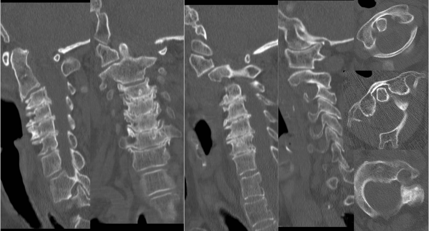
Sagittal, coronal, and axial CT images of the cervical spine demonstrate abnormally accentuated rightward rotation of C1 on C2. The left lateral mass of C1 is significantly subluxed anteriorly with respect to the lateral mass of C2. On one of the coronal MPR images, the odontoid proces abuts the right lateral mass of C1. There is also approximately 2 mm anterior subluxation at C2-3 and severe multilevel degenerative disc disease with osteophytes and discogenic sclerosis.
Discussion/Differential Diagnosis:
Any time there is a rotation of C1 on C2, the more common physiologic motion must be distinguished from the rare fixed rotatory subluxation of C1 on C2. Much confusion and controversy exists regarding the entity of rotatory C1-2 subluxation(ARF). An important clinical distinction is the presence of a fixed painful torticollis with "Cock robin" head position slightly flexed, cranial rotated, and head tilting contralateral to the rotation direction. Normal physiologic motion of C1-2 may be between 30-50 degrees with capture of lower cervical rotation occurring at 20-30 degrees. Slight lateral tilting of the head may facilitate rotation due to relaxing the alar ligament. ARF may be suspected on radiographs when there is significant asymmetric position of the odontoid process with respect to the lateral mass of C1, provided the radiographs are precisely positioned. The apprarent sizes of the C1 lateral masses on frontal projection will also be discrepant due to the rotation, with the contralateral side having a more squared appearance. ARF may be diagnosed on CT in the neutral position if the rotation is obvious and not accounted for by positioning and head rotation, but can be confirmed wiith CT performed with head rotation, which will demonstrate a fixed C1-2 relationship regardless of head position. Rotation toward the deformity may be facilitated while rotation the opposite way will be restricted. With extreme rotations, there is a risk for vertebral artery and/or cord injury.
Fielding and Hawkins Classification of ARF:
type 1- fixed ARF with intact transverse ligament
type 2- disruption of transverse ligament with 3-5 mm atlantoaxial subluxation
type 3- disruption of alar and transverse ligament with greater than 5 mm AA sublux
type 4- (very rare), both lateral masses of C1 displaced anteriorly.
This discussion was condensed from Resnick et. al. Diagnosis of Bone and Joint Disorders 4th ed. Saunders 2002, pp.2955-2958.
Other references for above discussion:
Fielding, JW and Hawkins RJ. Atlanto-axial Rotatory Subluxation. J Bone Joint Surg Am 59:37, 1977.
Rinaldi, et al. Computerized tomographic demonstration of rotational atlantoaxial fixation: Case report. J Neurosurg 50:115, 1979.
Jacobson G and Adler DC. Examination of the atlantoaxial joint following injury with particular emphasis on rotational subluxation. AJR 76:1081, 1956.
BACK TO
MAIN PAGE