Reversible Splenial Lesion
Findings:
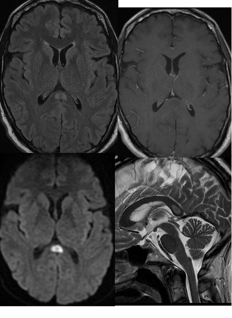
Axial FLAIR, axial T1 post contrast, sagittal T2, and axial diffusion weighted imaging demonstrates a well defined, symmetric, non-expansile, nonenhancing signal abnormality at midline involving the corpus callosum splenium. The splenium lesion demonstrates restricted diffusion.
Discussion/Differential Diagnosis:
The reversible splenial lesion is also known as MERS (mild encephalitis/encephalopathy with a reversible isolated SCC lesion), and is typically asymptomatic in isolation as opposed to other causes of corpus callosum lesions that will cause symptoms of hermispheric disconnection. These are classically associated with stopping or changing antiepileptic drugs, after a single seizure, after focal status epilepticus, but may also be seen with electrolyte imbalance, hypoglycemia, and uremia. The reversible splenial lesion is nearly always perfectly symmetric involving the central fibers, nonenhancing, and with DWI restriction. If the lesion does not fulfill these criteria, the wide range of other processes that may affect the corpus callosum should be considered such as MS/ADEM, PRES, DAI, Marchiafave Bignami disease, and neoplasm. The MERS lesion resolves within 1 month, most by 1 week.
BACK TO
MAIN PAGE