Left Vertebral Artery Pseudoaneurysm
Findings:
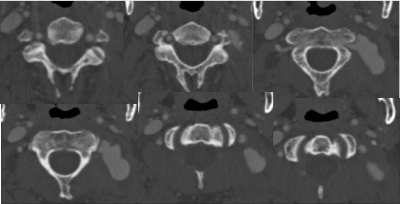
Axial CTA source images demonstrate an irregular large saccular projection from the posterior aspect of the left vertebral artery V3 segment.
Discussion/Differential Diagnosis:
The diagnosis in this case is easily made if you recognize that this enhancing lesion is directly contiguous with the arterial structure and has identical vascular enhancement. No specific history of trauma is noted, but the irregular morphology and location support that this is a pseudoaneurysm rather than a true saccular aneurysm. VA dissections may be extradural or intradural, with intradural dissections at a high risk for subarachnoid hemorrhage due to different vessel wall structure than the extracranial segments with less elastic fibers intracranially. VA dissections are most commonly from trauma including trivial sudden neck movement, but may be spontaneous in the circumstance of connective tissue disorders such as Ehlers Danlos and Marfan's. VA dissection occurs most commonly in the V2 and V3 segments with or without associated fractures.
Vertebral artery segments: V1- origin to transverse foramen entry, V2- intraforaminal through C2, V3- C2-C1, V4- intradural through VBJ
Dissection classification (Biffl): grade I- minor intimal injury w stenosis less than 25%, grade II- raised flap w >25% stenosis, intramural hematoma +/- intraluminal thrombus, grade III-pseudoaneurysm, grade IV- complete occlusion/thrombosis, grade V- transection
BACK TO
MAIN PAGE