Lymphoma
Findings:
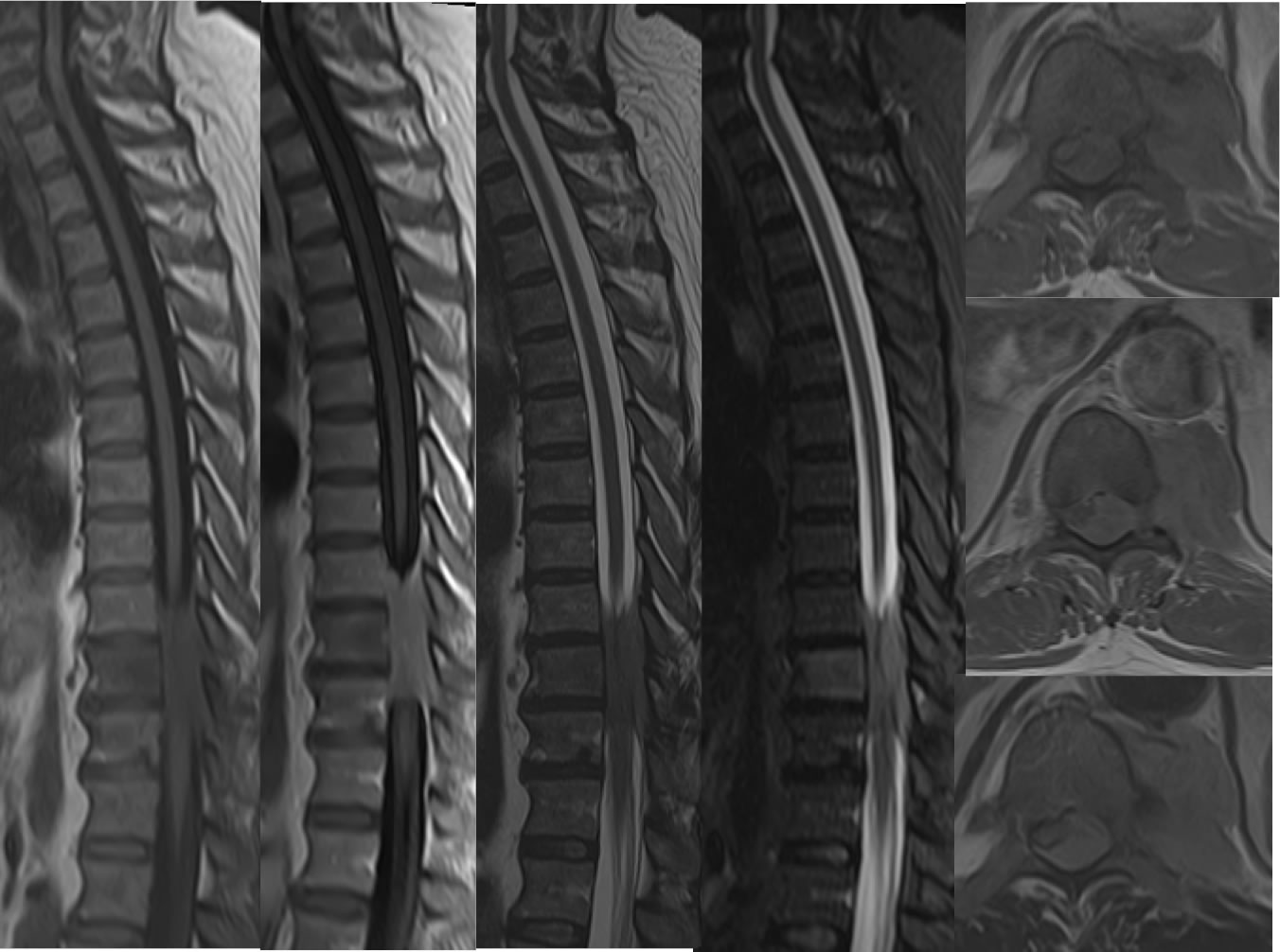
Sagittal T1, T1 post contrast, T2, and STIR images demonstrate a poorly defined infiltrative enhancing mass involving the epidural space nearly circumferentially, causing significant cord compression. There is adjacent abnormal marrow signal at T10. Axial post contrast T1 and T2 weighted images demonstrate the extent of the mass into the spinal canal causing cord compression.
Discussion/Differential Diagnosis:
The configuration of this mass with solid nearly homogenous nodular enhancement, infiltrative characteristics involving the spinal canal and ajacent osseous structures indicates a malignant process rather than a benign neural origin tumor or an infectious inflammatory process. The primary differential diagnostic considerations for this include lymphoma and metastatic disease. The homogeneity and infiltrataion rather than frank bone destruction raises the higher likelihood of lymphoma.
BACK TO
MAIN PAGE