sphenoid sinus polyps
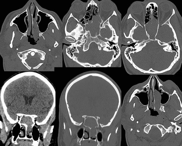
Findings:
Axial and coronal CT images through the paranasal sinuses, most with bone reconstructions, demonstrate complete opacification of the bilateral sphenoid sinuses with extent into the left posterior nasal cavity and nasopharynx. The opacification of the sphenoid sinuses is of soft tissue density. The bilateral sphenoid sinus ostia are widened. In some regions, the anterior sphenoid sinus walls are demineralized and destroyed. No significant expansile characteristic is identified. Scattered small air densities are present within the lobulated nasal and nasopharyngeal components.
Discussion/Differential Diagnosis:
The appearance of this process is nonspecific, and not optimally chracterized without MRI. The interface between obstructing lesion at the sphenoid ostium and postobstructive inflammatory disease is not distinct. The bubbly appearance of the component on the nasal cavity is more characteristic for benign polyps than malignancy. There is significant overlap in appearance between benign and malignant disease in this region, particularly with the anterior sphenoid wall destruction. Unless the process is of very low fluid density, the density of the lesion cannot be used as a distinguishing feature between benign and malignant disease.
BACK TO
MAIN PAGE