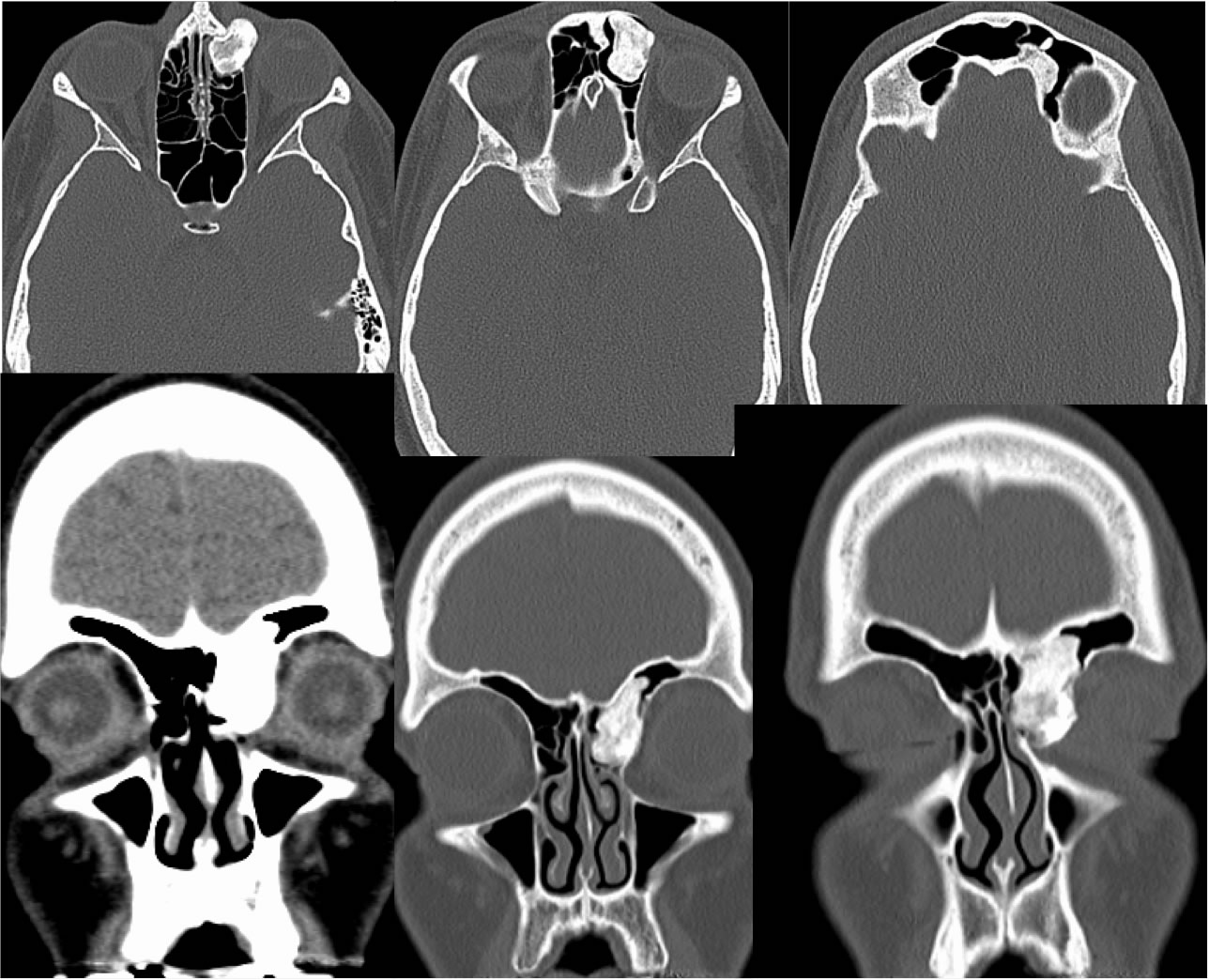
Fibroosseous Lesion Ethmoid
Findings:
Multiple axial and coronal CT sinus images, most with bone reconstructions, demonstrate a densely ossified expansile lesion near the left frontoethmoid junction, extending into the medial orbit and frontal sinus. There is no associated soft tissue mass or aggressive bone destruction.
Discussion/Differential Diagnosis:
Benign fibro-osseous lesions of the paranasal sinuses include fibrous dysplasia, ossifying fibroma, and osteoma in order of degree of ossified matrix. When small, these are typically asymptomatic and seen as incidental findings in up to 3% of sinus CT. The differential diagnosis includes other bone forming tumors such as osteoblastoma or osteosarcoma, but a more aggressive appearance would be expected. When large, they may become symptomatic due to obstructive or local pressure effects. Smaller osteomas may be symptomatic due to innervation and inflammatory mediators. Osteomas are most common in the frontal sinuses followed by ethmoid and are rare in sphenoid sinuses. Histologically, these are osteogenic tumors with mature bone, with or without a haversian system, and may contain marrow. Treatment options vary with physician, some may observe, with obstructing lesions usually resected.
BACK TO
MAIN PAGE