Neurofibromatosis Type 2
Findings:
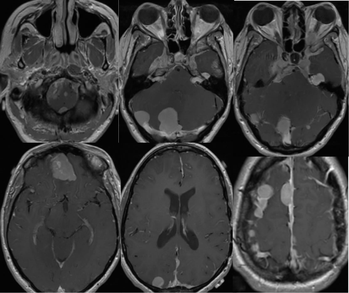
Multiple axial T1 weighted post contrast images demonstrate innumerable extra axial masses over both convexities and falx demonstrating homogeneous enhancement. Additional nodular masses are present at the foramen magnum, bilateral internal auditory canals, bilateral Meckels caves, left orbital apex, bilateral cavernous sinuses, and inferior right frontal region. Left parietal craniotomy is incompletely included. There is effacement of structures at foramen magnum magnum and there is distortion of the fourth ventricle, with no significant hydrocephalus visible. Some of the masses are inseparable from and likely invade dural sinuses.
Discussion/Differential Diagnosis:
The differential diagnosis for this process is limited, the presence of extensive extraaxial masses including bilateral neurogenic tumors indicated that NF2 is most likely. Metastatic disease can appear similar, but would not be typical to occur as a manifestation of mets from unknown primary in a young adult. Discussion and additional cases of NF2:
-Neurofibromatosis type 2
-Neurofibromatosis type 2(2)
BACK TO
MAIN PAGE