5FU toxicity
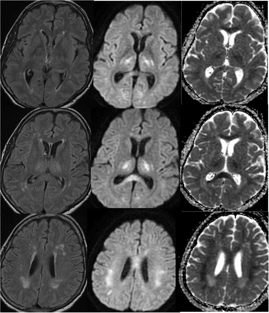
Findings:
Matched axial FLAIR, DWI, and ADC images demonstrate confluent nearly perfectly symmetric zones of signal abnormality and diffusion restriction within the bilateral thalami, corpus callosum, and deep parietal white matter. In some areas, FLAIR signal changes are absent or inconspicuous, while there is signal abnormality on diffusion and ADC map in these regions. Scattered superimposed patchy and punctate zones of white matter disease are present which are likely due to chronic microvascular ischemia.
Discussion/Differential Diagnosis:
The symmetry of the signal changes should raise suspicion for a metabolic or toxic process, which should cause a search of the medical history for clues.The differential diagnosis includes a toxic leukoencephalopathy (TLE), extrapontine myelinolysis, or hypoxic insult. This patient was undergoing 5-FU therapy for cancer. 5-FU is one of many agents known to cause TLE, including but not limited to antineoplastics, immunosuppressives, antibiotics, inhalants, illicit drugs, and CO poisoning. The clinical presentation is highly variable and nonspecific, with many minimally symptomatic cases not undergoing imaging. Improvement is expected after cessation of the underlying cause. Symmetric involvement of deep white matter and corpus callosum with restricted diffusion seems to be characteristic for 5-FU toxicity. An acute hyperammonemic TLE may occur after 5-FU therapy in the absence of imaging abnormalities.
BACK TO
MAIN PAGE