Labyrinthitis Ossificans
Findings:
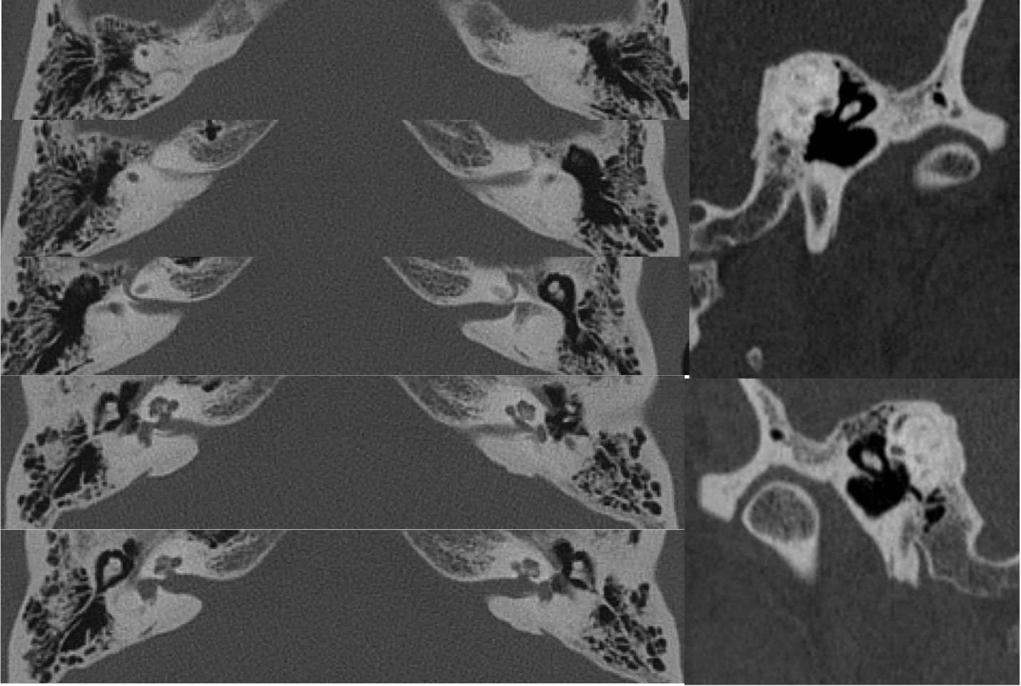
Axial temporal bone CT images with selected oblique modified reconstructions through the otic capsules demonstrate extensive osseous obliteration of the semicircular canals bilaterally. Additional ossifications are present with in the bilateral cochlea, left greater than right. The included mastoid air cells and middle ear cavities are clear. The ossicular structures appear normal.
Discussion/Differential Diagnosis:
Inner ear structures are still visible, so this is not a congenital aplasia, but labyrinthitis ossificans (LO) may simulate a congenital aplasia if there is rare solid osseous obliteration with no visible structure. Cochlear otosclerosis would be limited to the cochlea and more commonly with demineralized cochlear margins than true sclerosis. LO may range from subtle increased CT density within the labyrinth to dense obliterative ossification. LO occurs rarely as as healing reaction after any type of insult to the inner ear, most commonly infection, but may also be seen after trauma or surgery. LO may complicate cochlear implantation. Areas of signal dropout due to ossification may be seen on MR, including absence of the expected T2 hyperintensity of inner ear structures.
BACK TO
MAIN PAGE