Herpes Encephalitis
Findings:
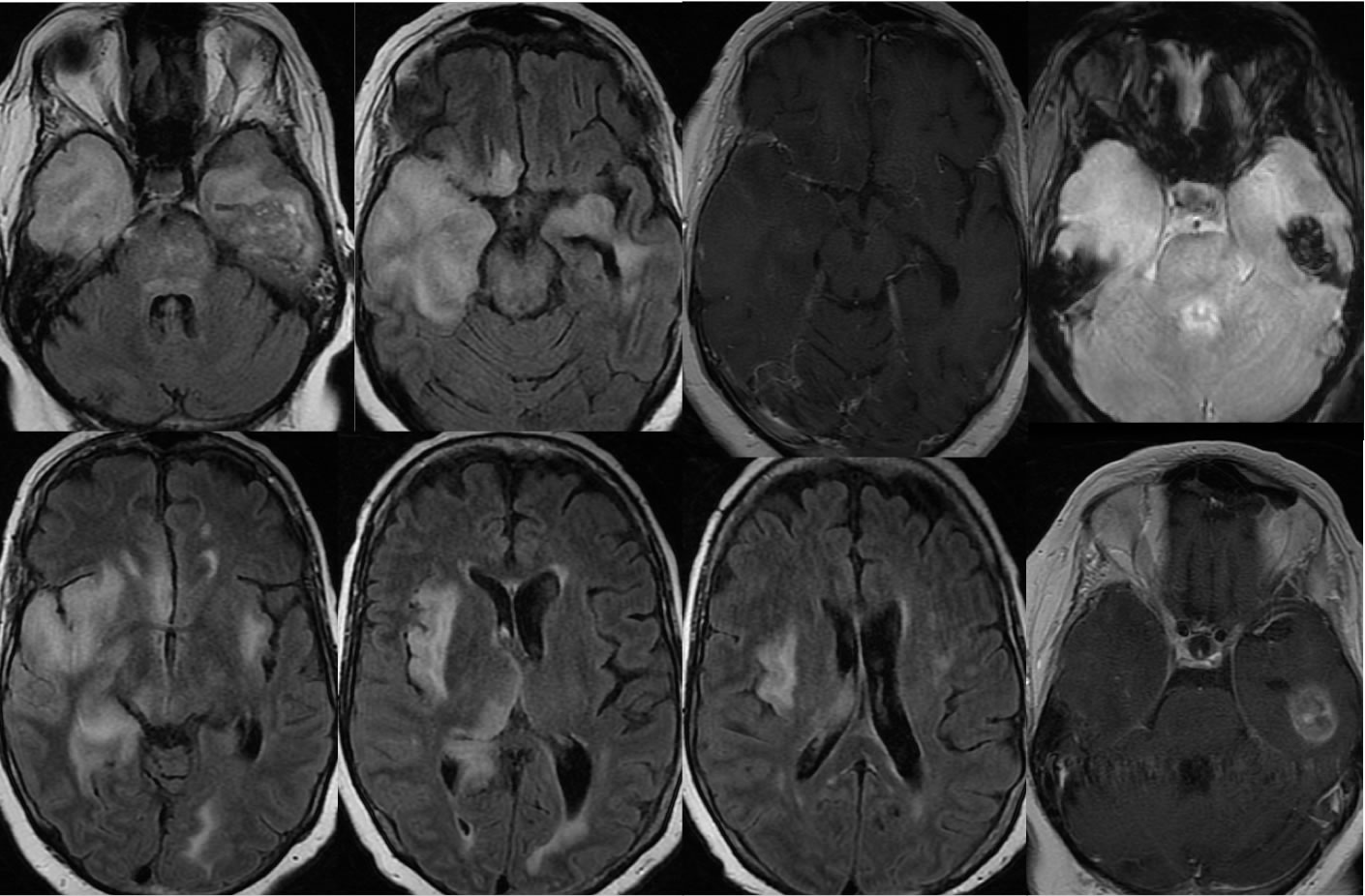
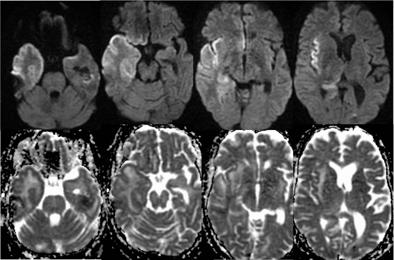
Multiple axial FLAIR images demonstrate bilateral temporal lobe signal abnormalities right much greater than left with mild mass effect on the right. The infiltrative signal abnormalities extend into the right hippocampus, right insula, and right thalamus. Minimal extent into the left subinsular region is noted. A few nonspecific superimposed white matter hyperintensities are present. The post contrast images demonstrate no abnormal enhancement of this process. The axial gradient echo image demonstrates an oval zone of hemorrhagic change within the right temporal lobe, with hyperintensity on the post contrast image in part likely representing methemoglobin blood products. Diffusion and ADC images demonstrate ribbon like zones of restricted diffusion involving the right frontotemporal and insular cortex also involving the right hippocampus. Susceptibility artifact is seen in the region of the temporal lobe hematoma.
Discussion/Differential Diagnosis:
The differential diagnosis for this process includes encephalitis, glial neoplasm, and infarction. The presence of bilateral temporal lobe signal abnormalities which involve the insular cortex and associated with diffusion restriction should raise suspicion for HSV encephalitis. Other cases and discussion of HSV encephalitis:
BACK TO
MAIN PAGE