Postictal Signal Changes
Findings:
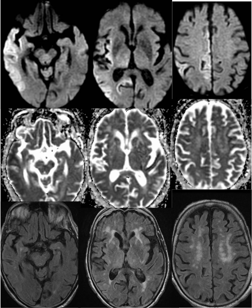
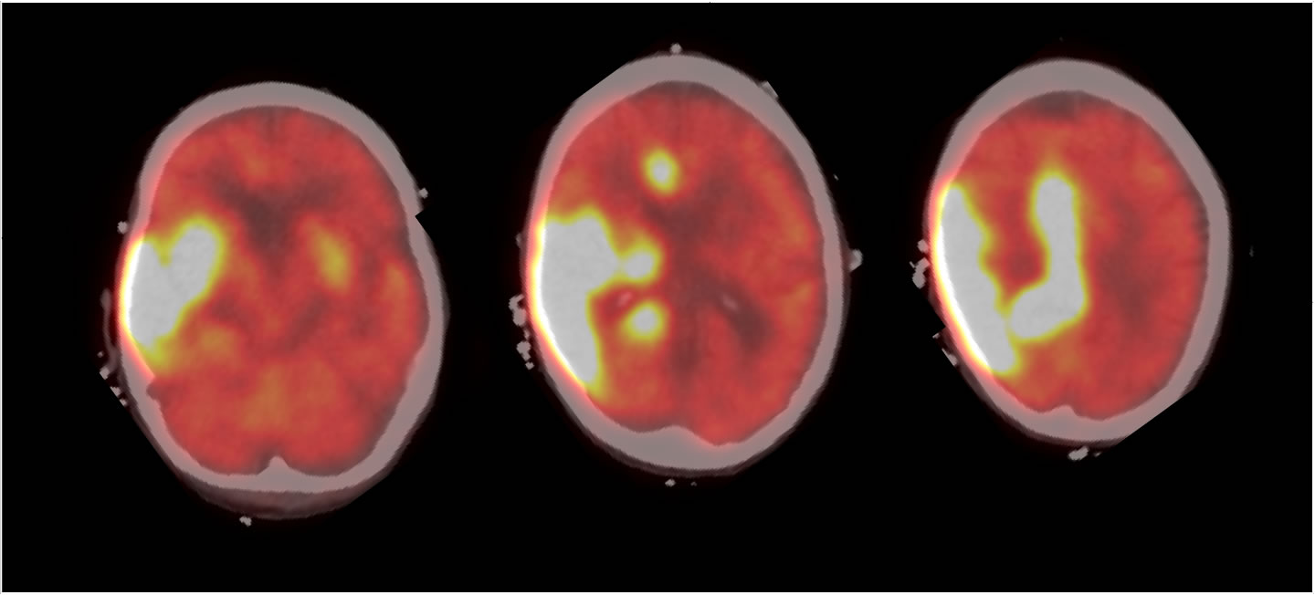
Axial diffusion and ADC map demonstrates restricted diffusion within the right temporal cortex, right frontal parasagittal cortex, and medial right parietal cortex, also involving the insula. Associated FLAIR signal alteration is inconspicuous. No mass effect or edema is seen. Superimposed chronic microvascular ischemic white matter changes are incidentally noted. PET brain imaging demonstrates intense hypermetabolism in the regions of diffusion abnormality.
Discussion/Differential Diagnosis:
The differential diagnosis includes seizure related signal abnormalities and infarction. The DWI signal incompletely involves multiple unilateral vascular distributions which would make multiple embolic infarction unlikely. While the precess does involve insula, cingulate gyrus, and the lateral temporal lobe like HSV encephalitis, the lack of edema, mass effect, or significant FLAIR hyperintensity would make HSV extremely unlikely. PET reflects the high metabolic demand of seizing brain. FLAIR hyperintensity, mild swelling, DWI restriction results from cortical cytotoxic edema. Enhancement on MR may be variable, sometimes very intense and gyriform simulating subacute infarction. These changes often resolve after cessation of seizure activity, but cortical infarction may occur with very prolonged status epilepticus.
BACK TO
MAIN PAGE