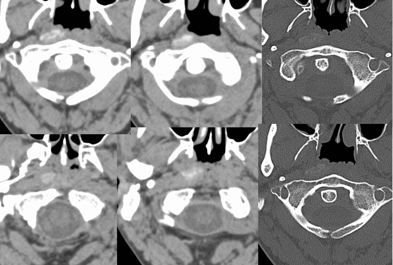
Calcific Longus Colli Tendinitis
Findings:
Axial noncontrast CT images demonstrate patchy hyperdensity of the right longus colli muscle near the C1 level. Congenital nonfusion posterior arch C1 is incidentally noted.
Discussion/Differential Diagnosis:
The presence of calcification within a muscle is typically a benign process including myositis ossificans that may result from any type of trauma with muscle rupture and/or hematoma that calcifies. There is no pathologic proof on this case and mysotis ossificans cannot be excluded, but the location of cacification is somewhat typical for calcific longus colli tendonitis (CLCT). CLCT is a type of hydroxyapatite deposition disease and is an inflammatory granulomatous reaction to the crystal deposition. Absent in this case are expected surrounding inflammatory edema and standing due to an active process. In the acute phase, patients may have severe neck pain that simulates more worrisome etiologies, however when this is appropriately diagnosed by CT, it responds to conservative therapy. It is a diagnosis rarely considered by clinicians and radiologists are critical in this pathway.
BACK TO
MAIN PAGE