Nasoethmoid Encephalocele
Findings:
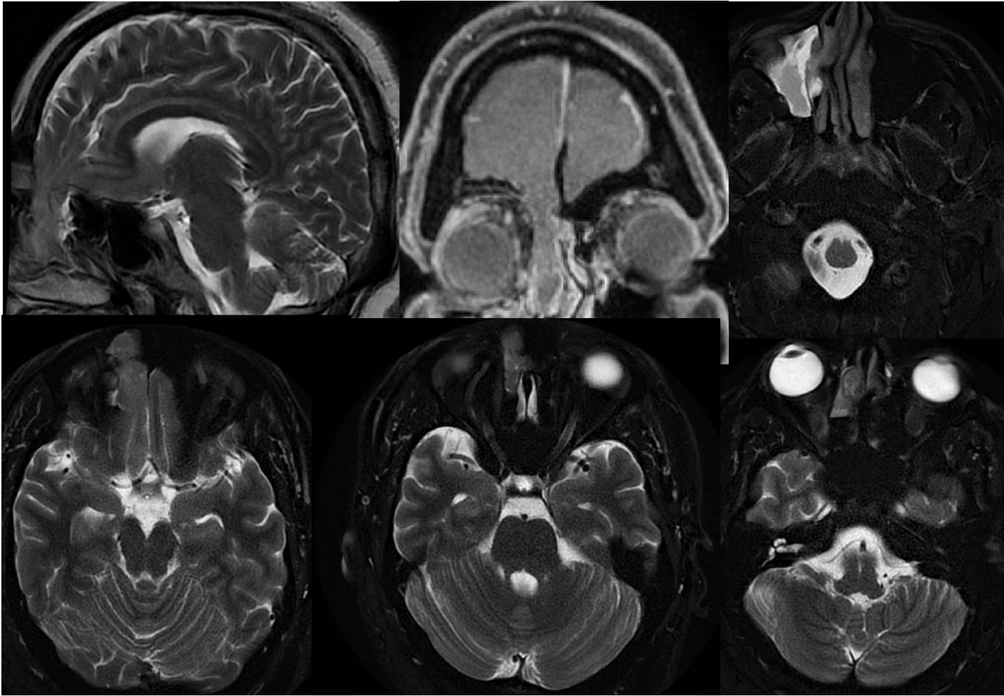
Multiple MR images demonstrate a large defect in the right cribriform plate, with significant inferior extent of the right inferior frontal lobe and meninges into the nasoethmoid cavity. Superimposed chronic appearing R maxillary sinus inflammatory disease is incompletely shown.
Discussion/Differential Diagnosis:
A nasoethmoid mass has a broad differential diagnosis including but not limited to inflammatory disease, polyp, mucocele, neoplasm, and encephalocele. The documentation of tissue following brain signal in this region is critical to diagnose an encephalocele, including the direct contiguity with the intracranial compartment. There is also the characteristic cascading appearance on the sagittal image. Imaging prior to intervention for a nasoethmoid mass is essential to avoid misdiagnosis and complications. The nasoethmoid encephaloceles are typically thought of as congenital anomalies, but in this case this is an adult with an acquired encephalocele herniating through a preexisting defect in the cribriform plate.
BACK TO
MAIN PAGE