Adenoid Cystic Carcinoma, Submandibular Gland
Findings:
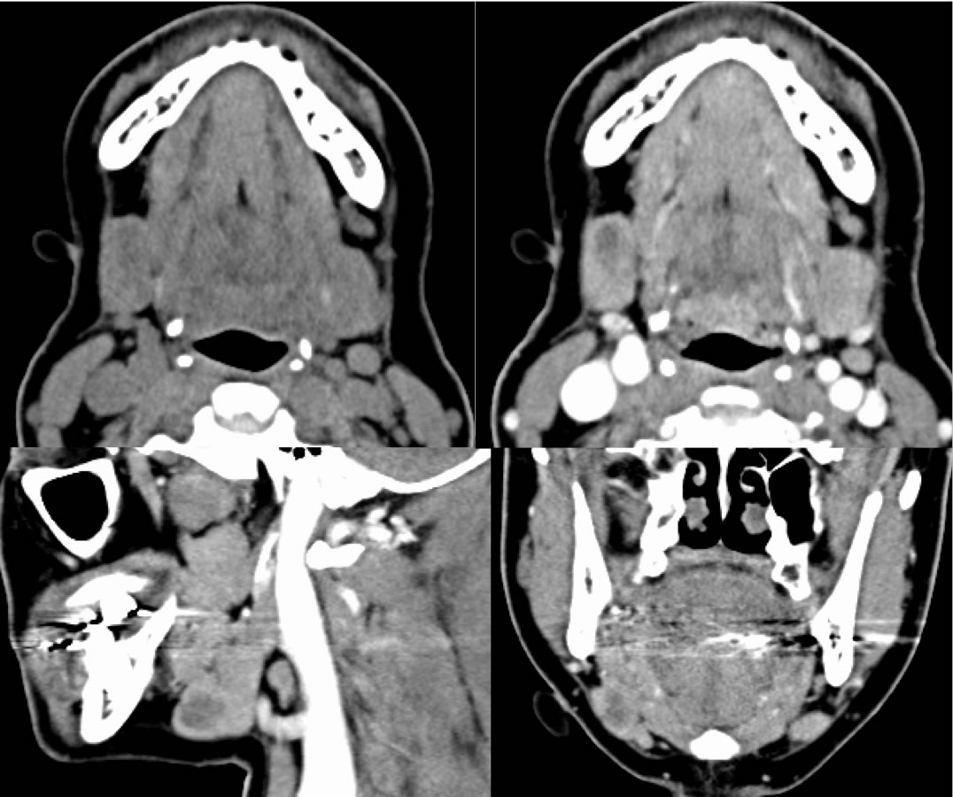
Pre-and post contrast neck CT images demonstrate a poorly defined focus of decreased attenuation with in the right submandibular gland. Of note, there is no surrounding inflammatory change, thickening of the platysma muscle, dilated ductal system, calculus, or well-defined enhancing wall present.
Discussion/Differential Diagnosis:
Similar to neoplastic disease seen in many other organs, a poorly defined zone of decreased attenuation within the structure in the absence of surrounding soft tissue stranding, abnormally dilated ductal system, and/or overall enlargement of the gland should raise suspicion for neoplasm rather than an inflammatory process. Adenoid cystic cancer (ACC)is rare, but is most common in the submandibular gland. It is the classic tumor to be associated with perineural tumor spread, although squamous cell being the most common head and neck cancer has the highest overall number of cases with perineural spread. ACC is one of the head and neck cancers known for a propensity for often late distant metastases including lungs, liver, bone, and brain, despite demonstrating low grade histology. ACC has a prolonged clinical course with poor overall long term survival. They may also occur as primary tumors in a wide variety of other sites, including lung, breast, other head and neck sites, and in the uterus. The imaging findings are nonspecific malignant pattern- solid, enhancing, poorly defined, mixed T2 cellular signal simlar to other malignant salivary gland neoplasms, however particular attention must be paid to known sites of perineural tumor spread, such as skull base foramina including foramen ovale and rotundum.
BACK TO
MAIN PAGE