Metastatic Breast Cancer
Findings:
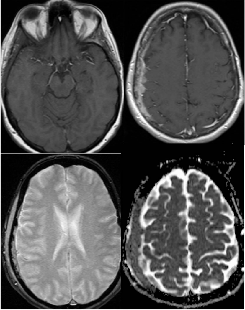
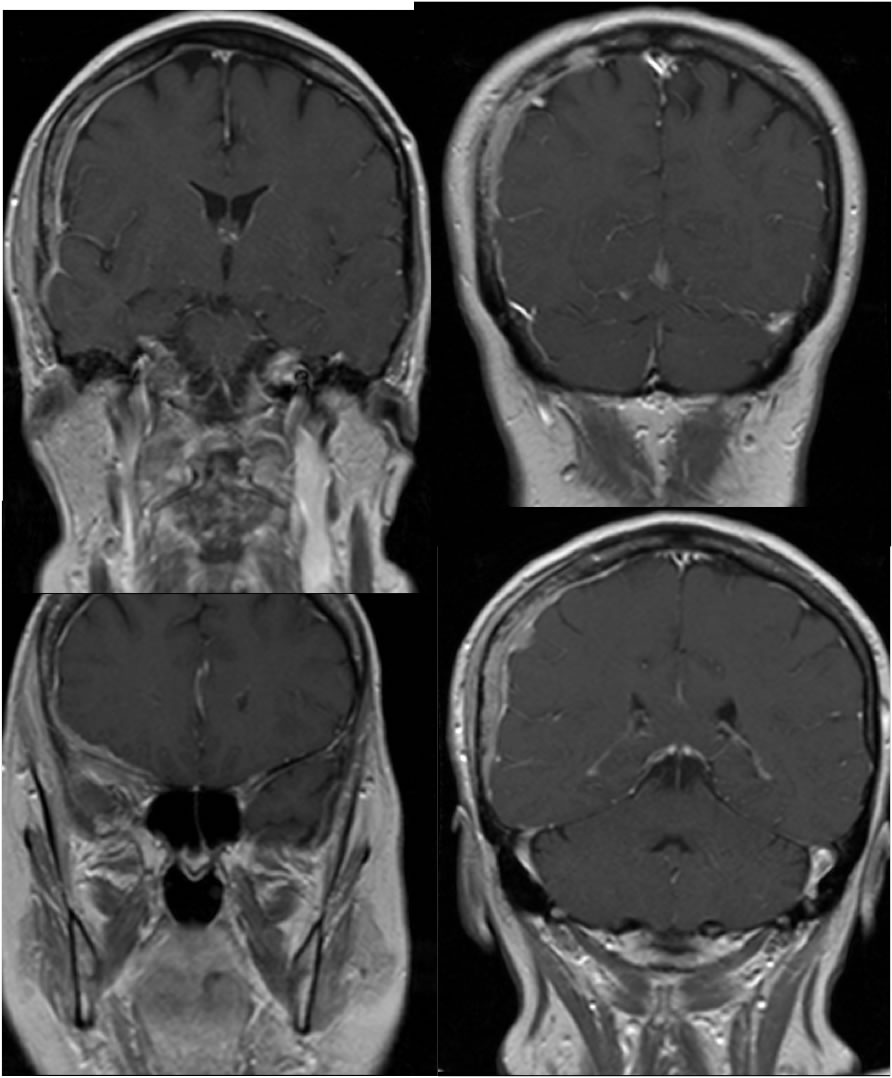
Multiple MR images demonstrate extensive marrow infiltration and abnormal enhancement of the right parietal calvarium with underlying irregular nodular dural thickening and enhancement. In some regions, nodular dural thickening is inseparable from underlying brain cortex. Overlying subgaleal soft tissue mass is present. The process demonstrates reduced ADC values. The T1 precontrast image demonstrates infiltration of the adjacent temporalis muscle and diffuse low signal intensity within the calvarial marrow including the sphenoid wing.
Discussion/Differential Diagnosis:
The primary differential diagnostic considerations for this lesion include metastatic disease, meningioma, and inflammatory process such as sarcoidosis. Sarcoidosis may be indistiguishable, clinical history may help, but in this case is less likely due to the primary bone involvement, secondary dural involvement, and subgaleal mass. While intraosseous meningiomas do occur, smooth expansion of bone would be expected. Although no CT is available, the appearance on coronal images suggests that there is permeative bone destruction, and the irregular dural thickening and nodularity confirms that metastases are most likely.
Bone is the most common site of breast cancer mets, followed by lung, liver, nodes, and brain. Symptoms are variable and may not correlate with the extent. Patients with isolated bone mets in the absence of other organ mets are typically younger and have an improved survival rate. (condensed from Steinauer, K; Huang, D; et al. Bone metastases in breast cancer: Frequency, metastatic pattern and non-systemic locoregional therapy. J Bone Oncol. 2014 May;3(2):54-60.)
BACK TO
MAIN PAGE