Frontotemporal Lobar Dementia
Findings:
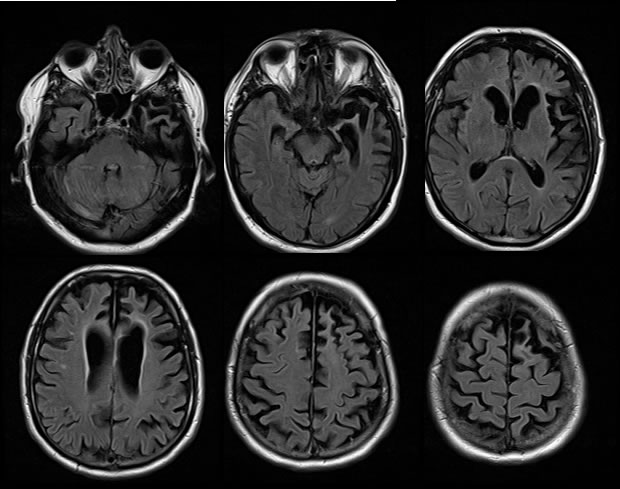
Axial FLAIR images demonstrate marked asymmetric atrophy of the left frontal and temporal lobes, with marked volume loss of the medial left temporal lobe. There is also severe atrophy involving the right frontal lobe, and mild to moderate atrophy involving the right medial temporal lobe. Subtle cortical gliosis is also visible in the regions of marked atrophy.
Discussion/Differential Diagnosis:
The differential diagnosis for atrophy is broad, with some helpful features including the pattern and symmetry of atrophy. Marked atrophy may be seen with any type of remote insult, and clinical history may help distinguish the etiology. In this case, concern should be raised for a neurodegenerative process including frontotemporal lobar degeneration and Alzheimers disease, but Alzheimers disease would not be expected to predominantly involve the frontal lobes.
Frontotemporal lobar degeneration (FTLD) used to be known as Picks disease, but this should be reserved for a pathologic diagnosis with Pick bodies visible. Variants of FTLD exist, including a behavioral variant which predominately involves frontal lobes, a language variant which involves temporal lobes left greater than right, with additional subtypes based on the type of aphasia. Most cases are sporadic, up to 40% may be autosomal, with a younger onset then Alzheimers disease. The behavioral variant is associated with more caudate atrophy. Asymmetric involvement is common.
BACK TO
MAIN PAGE