Superficial Siderosis
Findings:
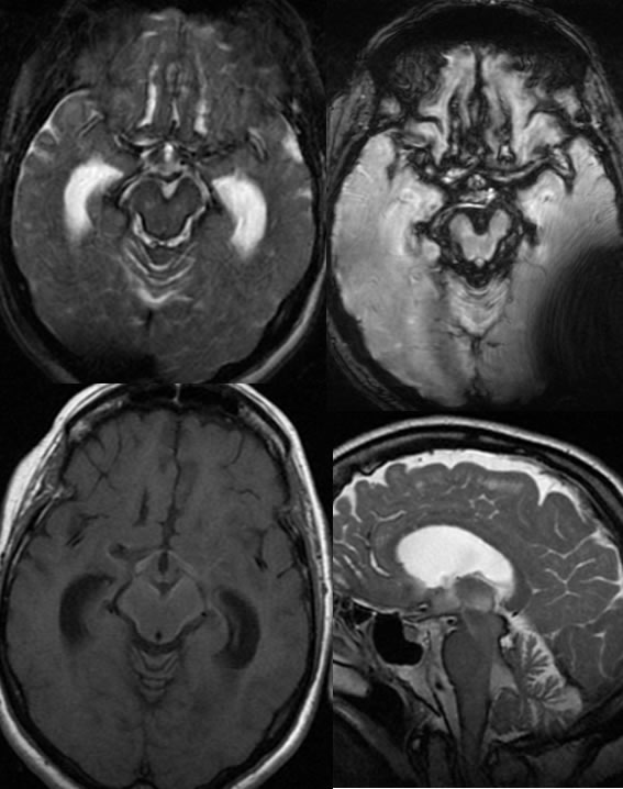
Multiple MR images demonstrate extensive T2 hypointense coating of the brainstem and subarachnoid spaces extending along the sylvian fissures and the cerebellum. T1 weighted imaging demonstrates hyperintensity in these regions. There is associated cerebellar and brainstem atrophy. The included ventricular system is dilated.
Discussion/Differential Diagnosis:
Superficial siderosis has no other reasonable differential diagnostic possibility, but can be caused by a wide variety of processes. Any type of remote and typically repeated subarachnoid hemorrhage can cause siderosis. Other cases of superficial siderosis:
-superficial
siderosis1
-superficial
siderosis2
BACK TO
MAIN PAGE