Congenital Unilateral Perisylvian Syndrome
Findings:
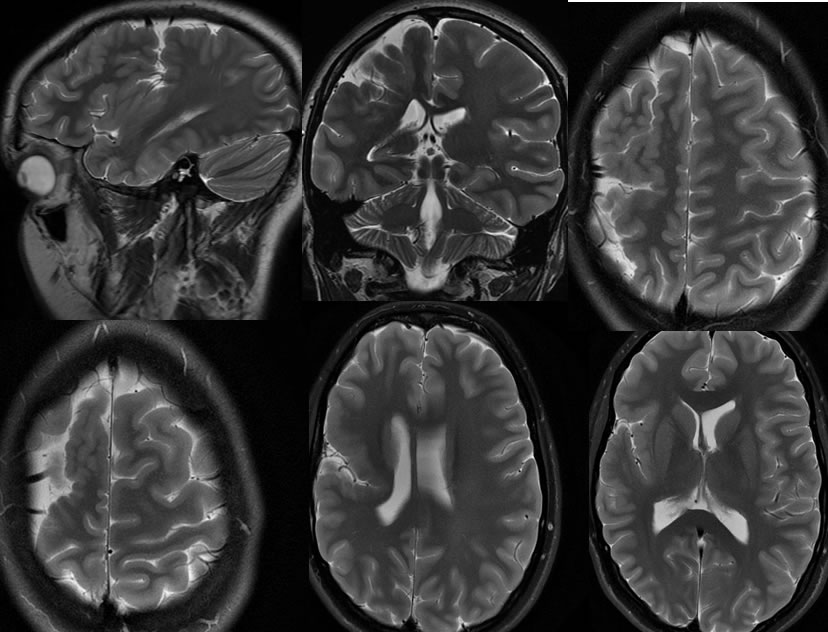
Multiplanar T2 weighted imaging demonstrates asymmetric volume loss of the right cerebral hemisphere. The right sylvian fissure is unusually deep and vertically oriented, with abnormal sulcation lining the deep cleft. Zones of polymicrogyria are seen with other areas of cortical thickening due to cortical dysplasia. The right lateral ventricle is not asymmetrically dilated. A thin white matter band is seen in the right corona radiata along the depth of the abnormal right sylvian fissure.
Discussion/Differential Diagnosis:
A deep brain cleft often raises suspicion for schizencephaly, however the dimpling of the ventricular margin is not visible in this case, and gray matter does not extend all the way to the ventricular margin, which must be present with schizencephaly whether open lipped or closed lipped.
Perisylvian syndrome is a very rare late migrational cortical organizaton anomaly that is most commonly bilateral and associated with characteristic features of an unusually deep and vertically oriented sylvian fissure that is lined by abnormal cortex. Polymicrogyria and thick cortical dysplasia lines the cleft. Clinically, these patients have seizures, and cognitive impairment, and pseudobulbar palsy. The unilateral form is associated with contralateral hemiparesis. As expected, the clinical sequelae are far more prominent with bilateral than unilateral involvement.
BACK TO
MAIN PAGE