Posterolateral Epidural Disc Extrusion
Findings:
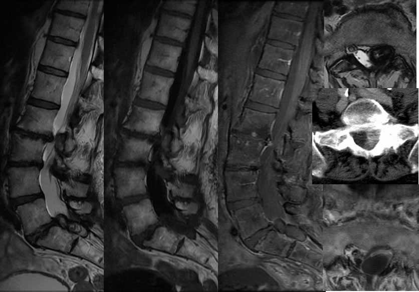
Multiple MR images demonstrate severe multilevel disc dessication and height loss with osteophytes and discogenic endplate signal alterations. At the L5-S1 level, there is a large septated appearing lesion in the left lateral and posterior epidural space, which causes mild thecal sac compression and displacement/compression of the left S1 nerve root. The lesion is centrally T2 hyperintense with a markedly hypointense rim, does not enhance, and is partially attached to the
Discussion/Differential Diagnosis:
The only reasonable diagnostic considerations for this process are a posterolateral epidural disc extrusion and a synovial cyst. Enhancement would be expected with neoplastic disease. Rim enhancement would be expected with a synovial cyst, therefore a posterolateral disc should be considered first. In addition, the central contents of the lesion follow the signal and attenuation of disc material. A connection to the parent disc may be confirmatory. This type of disc extruson is exceedingly rare, with only a few case reports available. The process becomes more of a diagnostic dilemma when the disc is sequestered, at this point it may show peripheral enhancement and simulate other causes of epidural masses.
BACK TO
MAIN PAGE