Acute Left Parotitis
Findings:
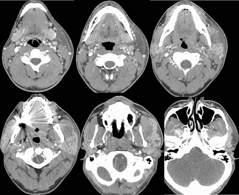
Multiple contrast enhanced neck CT images are presented. The left parotid gland is enlarged and demonstrates asymmetric enhancement with no focal mass. There is extensive surrounding soft tissue induration which extends into the left submandibular region. The left platysma muscle is asymmetrically thickened. No dilated ductal system or radiopaque calculi are seen.
Differential Diagnosis:
Parotitis, infiltrative salivary gland tumor, trauma.
Discussion:
Acute bacterial parotitis is typically unilateral and manifests as diffuse parotid swelling and pain in a debilitated patient, caused by staph aureus in up to 90%. The parotid gland is by far the most common site of salivary gland infection. A careful search should be performed for calculi, dilated ducts, or underlying mass or fluid collection. Comorbidities such as dehydration, poor oral hygiene, malnutrition, reduced salivary output of many causes, calculi, and immunosuppression predispose to this process. It typically responds well to antibiotics and drainage of any abscesses, but mortality is still substantial up to 20% due to the typical poor underlying medical condition of the patient. Bilateral parotiditis is typically viral, most commonly mumps but other viruses have been causative, and is usually self limited. Follow up imaging should be considered for any residual mass after the acute process has resolved.