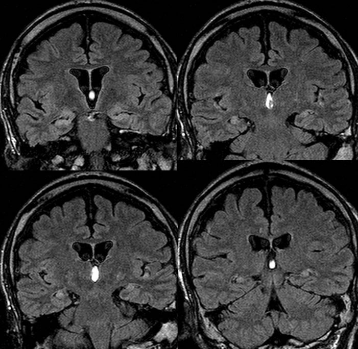

Mesial Temporal Sclerosis, Bilateral
Findings:
Coronal FLAIR images demonstrate volume loss and hyperintensity of the bilateral hippocampal formations.
Differential Diagnosis:
Bilateral MTS, remote infarcts, remote infectious/inflammatory
Discussion:
Mesial temporal sclerosis is a primary cause of partial complex seizures and is of uncertain underlying etiology, and may occasionally be seen in asymptomatic patients. It is thought to be either a developmental process, acquired from prolonged febrile seizures, status epilepticus, prolonged delivery, or ischemia, versus some combination of these. It is the most common indication for epilepsy surgery and a superimposed brain anomaly may be seen in 15%. Loss of hippocampal neurons and development of gliosis is seen histologically, most commonly involving the cornu ammonis. The classic imaging manifestations include volume loss and abnormal T2 hyperintensity of the hippocampus with loss of internal laminated architecture. Secondary findings include volume loss of other limbic structures including the fornix and mammillary body, as well as volume loss of temporal white matter tracts associated with asymmetric temporal horn. 20% may be bilateral. It is best detected on high resolution thin section MR images through the temporal lobes that are perpendicular to the orientation of the hippocampus. An expansile appearance should suggest an alternate diagnosis such as neoplasm or encephalitis. Tiny simple cysts within the hippocampi are typically symmetric and represent normal developmental variations as tiny inclusion cysts. The presence of MTS should prompt a careful search for other anomalies.