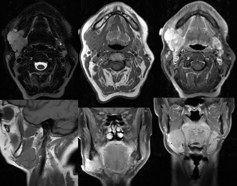
Lymphoma, R submandibular gland
Findings:
Multiple MR images demonstrate a homogeneously enhancing mass inseparable from the right submandibular gland. Normal submandibular gland tissue is displaced inferiorly and laterally, but the mass also infiltrates laterally where it is palpable. It shows intermediate signal on T-2 weighted imaging and is similar to slightly hyperintense to muscle on T1 weighted imaging.
Differential Diagnosis:
Lymphoma, pleomorphic adenoma, adenoid cystic carcinoma
Discussion:
The imaging appearance of this lesion is somewhat nonspecific, but the homogenous appearance should raise the possibility of lymphoma. Other types of salivary gland tumors in the above differential are typically more heterogenous in appearance, but there is some overlap. Primary lymphoma involving the salivary glands is rare, with 80% occurring in the parotid and 20% in the submandibular gland. They are considered MALT-omas, as tumors arising from mucosal associated lymphoid tissue along the GI tract. The prognosis for these lesions is reasonable with up to 50% disease free at 5 years. Secondary lymphoma involving the salivary glands is more rare and is usually large cell type with a poorer prognosis. Hodgkin’s lymphoma involving the parotid gland is uncommon, and the involved nodes are typically more heterogenous.