Subacute Hemorrhagic Contusions
Findings:
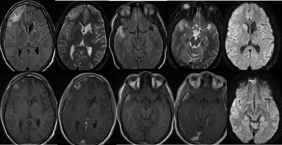
Multiple MR images demonstrate irregular signal abnormalities involving the periphery of the right frontal and temporal lobes . These lesions are not associated with restricted diffusion. Mild surrounding edema is present. The lesions demonstrate irregular peripheral enhancement and central zones of T1 precontrast hyperintensity compatible with hemorrhage. A remote left caudate infarct with volume loss is incidentally noted.
Differential Diagnosis:
Subacute contusions, abscesses, metastases.
Discussion:
Brain contusions are most commonly seen along peripheral aspects of the frontal and temporal lobes, often involving the orbital surfaces of the frontal lobes, anterior margins of the temporal lobes, and petrous apical surfaces of the temporal lobes where traumatic impact against bone is expected to occur. Occasionally these may occur opposite to the site of trauma as a contrecoup lesion. While this case simulates the appearance of other causes in the differential, the location is critical for correct diagnosis, and a history of head trauma was present. DWI failed to raise the possibility of abscess, and the patient also has no known primary cancer. Peripheral enhancement of these lesions is expected due to disruption of the blood brain barrier from injury.