Intramedullary Lipoma
Findings:
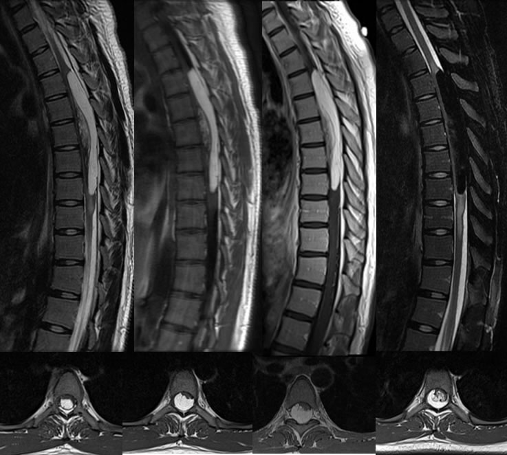
Multiple MR images demonstrate a very large intramedullary mass at midthoracic levels focally expanding the thoracic canal. Cord edema signal is seen along the superior and inferior margins of the lesion. In the midportion of the lesion, spinal cord fibers are splayed around an infiltrative component. The mass closely follows fat signal on all sequences, with subtotal suppression of signal on fat saturation. The post contrast images demonstrate no abnormal enhancement.
Differential Diagnosis:
Intramedullary lipoma, intradural extramedullary lipoma, liposarcoma, dermoid.
Discussion:
Intradural extramedullary lipomas (IDEML) are far more common than intramedullary lipomas although some may be subpial. Over 80% of IDEML are related to spinal dysraphism or are associated with the filum terminale, with other types of intradural lipomas being less than 1% of primary intraspinal tumors. The infiltrative appearance between fibers of the cord indicates that this is a very rare intramedullary lesion. These can be treated by surgical resection, with a high complication rate, therefore observation may be more appropriate if there is not prominent symptomatology. Since these are comprised of normal fat cells, they often shrink with weight loss particularly in obese patients, but this patient was not obese.