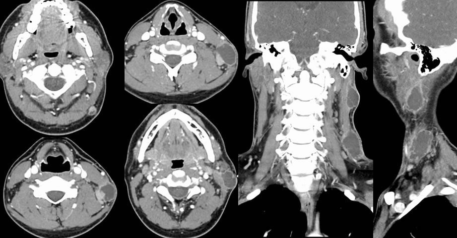

Bacterial neck abscesses, suppurative adenitis
Findings:
Contrast enhanced neck CT images demonstrate irregular peripherally enhancing subcutaneous lesions along the left neck. These lesions demonstrate central complex fluid attenuation and irregular enhancing walls. There is some more prominent enhancing tissue along the medial margin of the lesion near C6.
Differential Diagnosis:
Neck abscesses, necrotic malignant adenopathy, infected sebaceous cysts
Discussion:
The differential diagnosis of irregular rim enhancing lesions in the neck is fairly broad and clinical history is often critical. This patient had acute to subacute symptomatology with pain and redness at the sites as well as low grade fevers, no definite preexisting lesion. These were effectively cured with antibiotics with incision and drainage. Multifocal bilateral suppurative adenopathy with a more subacute course and little or no symptomatology should raise the possibility of more atypical organisms such as TB particularly if there is superimposed lung disease. Multifocal painless necrotic lymph nodes in an adult over 40 should raise the possibility of nodal metastases from a head and neck primary, and would be unusual as a manifestation of unknown primary. Common organisms responsible include staph and strep, with a head and neck source such as pharyngitis, sinusitis, or dental disease.