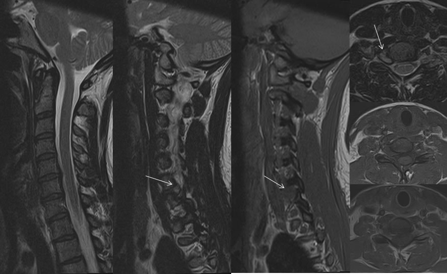
Right C6-7 foraminal soft disc herniation
Findings:
Sagittal and axial T-1 and T2 weighted images demonstrate a hyperintense lesion within the right C7 foramen causing severe narrowing of the right C7 foramen. There is no abnormal enhancement after gadolinium administration. The signal of the structure is slightly hyperintense to disc material, but it is contiguous with the disc margin.
Differential Diagnosis:
Herniated disc, neural origin tumor, metastasis
Discussion:
The absence of enhancement in this case effectively eliminates the possibility of tumor. Soft disc foraminal herniations are often less obvious than this and may follow nearly CSF signal on T2, only detectable by the mass effect on the exiting nerve root and displacement of the dural margin. The sagittal T1 imaging and axial T1 if performed is often very helpful for detection of soft discs, despite the oblique exit of the foramina seen on the sagittals. The soft disc will efface the normal perineural foraminal fat on T1.