Grade 3 malignant (anaplastic) meningioma
Findings:
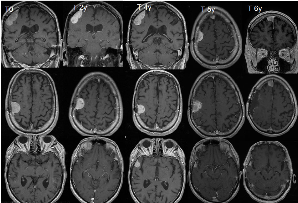
A series of T1 post contrast coronal and axial images performed over several years demonstrates postoperative changes of right parietal craniotomy with a homogeneously enhancing extra axial mass along the posterior margin of the craniotomy flap. An additional extraaxial mass over the right temporal convexity is seen to enlarge over four years. On the five-year scan, radiotherapy related changes are noted in association with the right frontal and right temporal extraaxial masses, but a new mass has developed in the anterior right parafalcine region. On the six-year scan, the right frontal extraaxial mass has been resected but the anterior right parafalcine lesion has enlarged.
Differential Diagnosis:
Malignant or atypical meningioma, metastases, meningeal sarcoma, postoperative dural thickening
Discussion:
The nodularity of these lesions and progression over time make postoperative dural thickening unlikely. Malignant meningiomas have typically aggressive features including indistinct margins, direct brain invasion, bone destruction, may penetrate extracranially, and have extensive vasogenic edema. While the individual lesions in this case do not appear particularly aggressive, the multiple recurrences should suggest a higher grade histology than the typical grade 1 meningioma.