Meningioma
Findings:
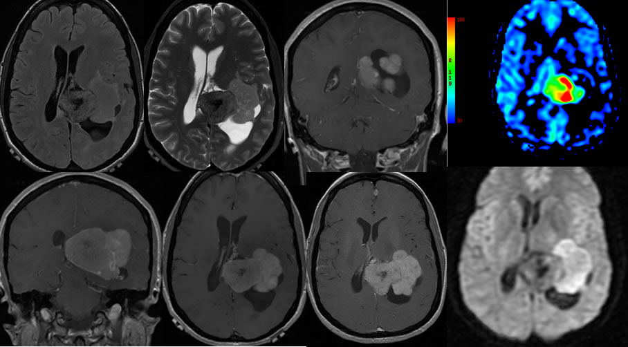
There is a large bilobedintraventricular mass centered within the posterior horn of the left lateral ventricle with associated dilation of the posterior horn and atrium. The medial component of the mass demonstrates central T1 and T2 hypointensity likely calcification. The peripheral aspect of the medial lobe is isointense to white matter on T1 and T2-weighted images. The lateral component of the mass is isointense to the gray matter on T2-weighted sequencesand shows restricted diffusion. The ASL perfusion image shows increased flow to the medial lobule of the mass. An additional small extraaxial mass projects over the anterior falx.
Discussion/Differential Diagnosis:
In this case, there appears to be two different histologies within the intraventricular mass, or this could represent two separate lesions that have become confluent over time. Multiple meningiomas are not uncommon, and should raise the suspicion of underlying NF2. The presence of restricted diffusion in meningiomas generally correlates with higher grade histology and more aggressive behavior, but the medial component shows much greater perfusion. Additional discussion of meningiomas is found here.
This case was in part prepared for presentation by Josh Hall, UC undergraduate.
BACK TO
MAIN PAGE