Colloid Cyst
Findings:
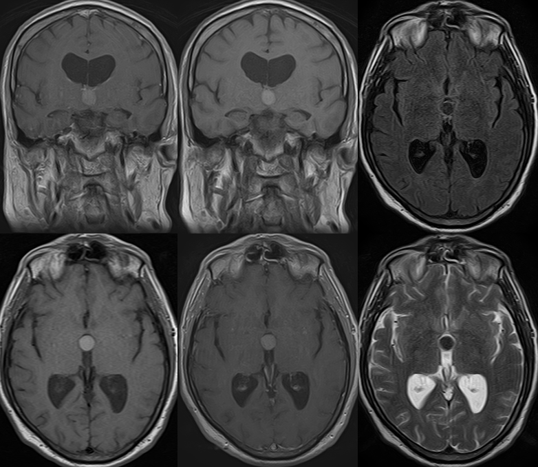
There is a well-circumscribed round lesion in the anterior third ventricle, slightly increased in signal on T1-weighted images, and decreased on T2 weighted sequences. It is thin-walled, and demonstrates no significant enhancement following contrast administration. It measures 1.4 x 1.4 cm in AP and transverse dimensions, by 1.6 cm in vertical diameter. It lies adjacent to the foramen of Monro bilaterally. There is some mild, symmetrical dilatation of the lateral ventricles bilaterally.
Discussion/Differential Diagnosis:
Differential Diagnosis: Colloid cyst, CSF flow artifact (confirm lesion in multiple planes), neoplasm such as subependymoma, craniopharyngioma, choroid plexus mass (expect enhancement).
-arise from endodermal inclusion, not ectoderm, similar to Rathke' s cleft cyst and foregut inclusion cysts
-epithelial lined, accumulate desquamed cellular debris and secretions (but less than 10% enlarge)
-typically resected through endoscopic approach w rare recurrence.
For additional discussion and examples, please refer to: Colloid cyst 1 and Colloid Cyst and Meningioma
This case was prepared with the assistance of Joshua Hall, UC undergraduate
BACK TO
MAIN PAGE