Intracranial Hypotension
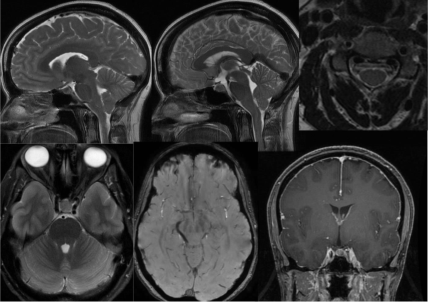
Findings:
Multiple MR images demonstrate inferior extent of the cerebellar tonsils below foramen magnum to the level of C1. There is inferior displacement of the brainstem with triangular pontine deformity. The pituitary gland is prominent. There is congestion of the cervical ventral epidural plexus. The dural sinuses are a distended with a rounded cross-sectional configuration. Minimally prominent linear dural enhancement is present over both cerebral convexities. CSF space in the optic nerve sheaths is not visible.
Differential Diagnosis:
The constellation of findings discussed above is highly suggestive of low CSF pressure. There is some overlap in imaging findings scene with high and low CSF pressure, for example there may be inferior displacement of the brainstem and cerebellar displacement in the presence of elevated CSF pressure. In the presence of into your brain stem displacement and elevated CSF pressure, in which case increased distention of CSF spaces over the convexities is often observed. The distention of dural sinuses rather than effacement indicates a high likelihood of low CSF pressure due to the inverse relationship of dural sinus distention and CSF pressure. Intracranial hypertension is sometimes associated with effacement of the bilateral transverse sigmoid sinus junctions. Care should be taken to distinguish abnormalities of CSF pressure from congenital anomalies such as Chiari malformation, since the treatment is different. When inferior cerebellar tonsilar displacement is seen, Chiari malformation cannot be assumed, and there should be a search for other findings that indicate an abnormality of CSF pressure. The absence of CSF space within optic nerve sheaths has been reported as a sensitive factor for detection of low CSF pressure.
Discussion:
Additional cases and discussion of CSF pressure issues:
-intracranial
hypotension
-spontaneous intracranial hypotension
-idiopathic intracranial
hypertension
BACK TO
MAIN PAGE