
Listeria Meningitis with Vasculitis
Findings:
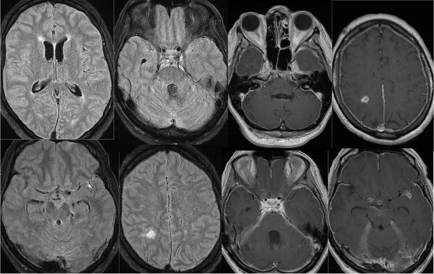
Multiple MR images demonstrate abnormal hyperintensity of the CSF spaces on FLAIR imaging, including leptomeningeal flair hyperintensity. There is relatively smooth pachymeningeal enhancement over both convexities come which also involves leptomeninges. Pachymeningeal enhancement extends into both internal auditory canals right greater than left and also lines the posterior fossa. A small enhancing lesion is seen in the right parietal lobe and there is irregular enhancement in the stem of the left sylvian fissure. The lateral ventricles are borderline prominent. Diffusion weighted image demonstrates foci of restricted diffusion compatible with infarcts in the left insula, right external capsule, and right occipital white matter. MRA images demonstrate multifocal vascular luminal irregularity with high-grade multifocal stenosis of the right MCA, high grade stenosis of the bilateral anterior cerebral arteries, and mild to moderate luminal regularity of the left MCA branches. There is mild luminal irregularity of the right posterior cerebral artery.
Differential Diagnosis/Discussion:
The differential diagnosis of leptomeningeal FLAIR hyperintensity includes hemorrhage, infectious/inflammatory, and metastatic neoplasm. Meningitis can cause an inflammatory vasculitis with associated infarcts. Other examples and discussion of meningitis:
-bacterial meningitis
-meningoencephalitis
BACK TO
MAIN PAGE