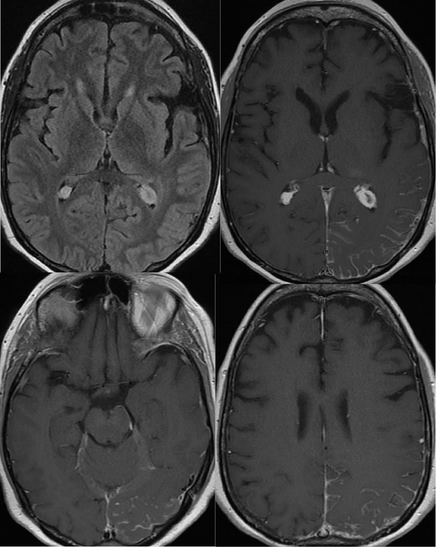
Pial Angiomatosis, Sturge Weber Variant
Findings:
Fine uniform linear leptomeningeal enhancement covers the left parietooccipital lobe, which is not associated with definite significant white matter FLAIR hypointensity underlying. There is enlargement and increased enhancement of the ipsilateral choroid plexus.
Discussion/Differential Diagnosis:
Leptomeningeal enhancement has a broad differential diagnosis, including meningitis, carcinomatosis, and granulomatous disease. Active abnormal leptomeningeal processes are often associated with subcortical T2 white matter hypointensity that is absent in this case. When enhancing leptomeninges are associated with ipsilateral choroid plexus enlargement in the absence of mass, a pial angiomatosis as seem with Sturge Weber syndrome should be considered. Volume loss and gyral calcification on CT are more characteristic for Sturge Weber. Additional discussion and cases of Sturge Weber below:
BACK TO
MAIN PAGE

