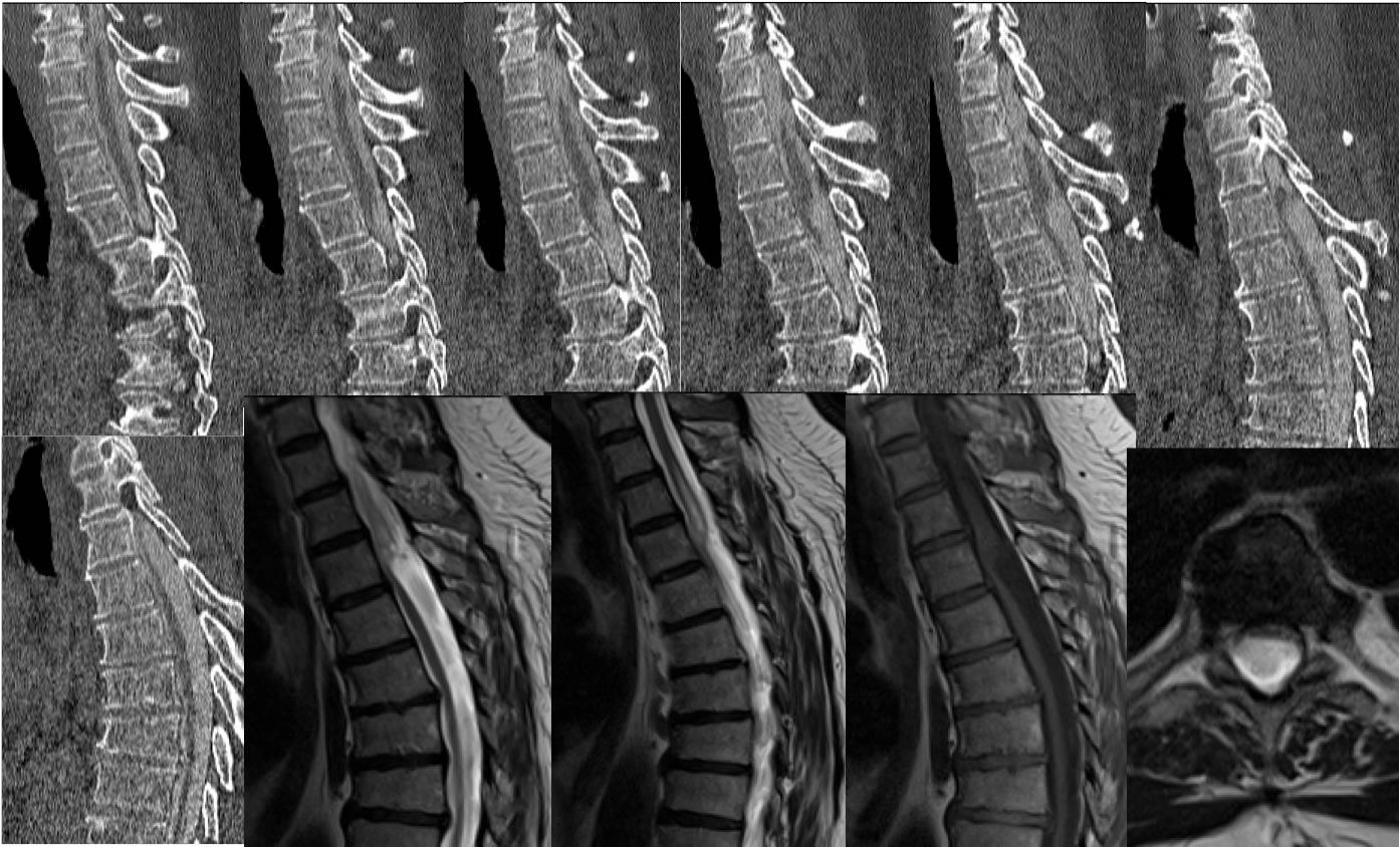
Ventral Cord Herniation
Findings:
Multiple sagittal CT myelogram images, sagittal T2, sagittal T1, and axial T2 weighted images demonstrate abrupt ventral deviation of the spinal cord near the T4-5 level which is inseparable from the posterior margin of T4. Patchy signal alteration is present within the spinal cord above this level without a well-defined syrinx visible. There is no cord expansion or cord signal abnormality below this level, but the cord does appear atrophic below T5. Marked flattening of spinal cord parenchyma as seen on the axial image. Superimposed degenerative disc disease is incidentally noted.
Discussion/Differential Diagnosis:
The differential diagnosis of abrupt ventral cord deviation is relatively narrow, and includes a dorsal arachnoid cyst causing ventral deviation of the cord, a focal adhesion, or ventral cord herniation. Additional case with discussion:
-ventral cord herniation
BACK TO
MAIN PAGE