Secondary Hyperparathyroidism (Lupus nephritis)
Findings:
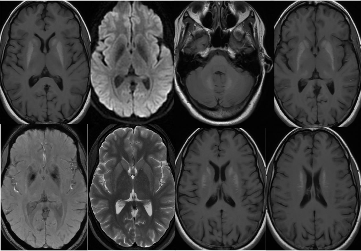
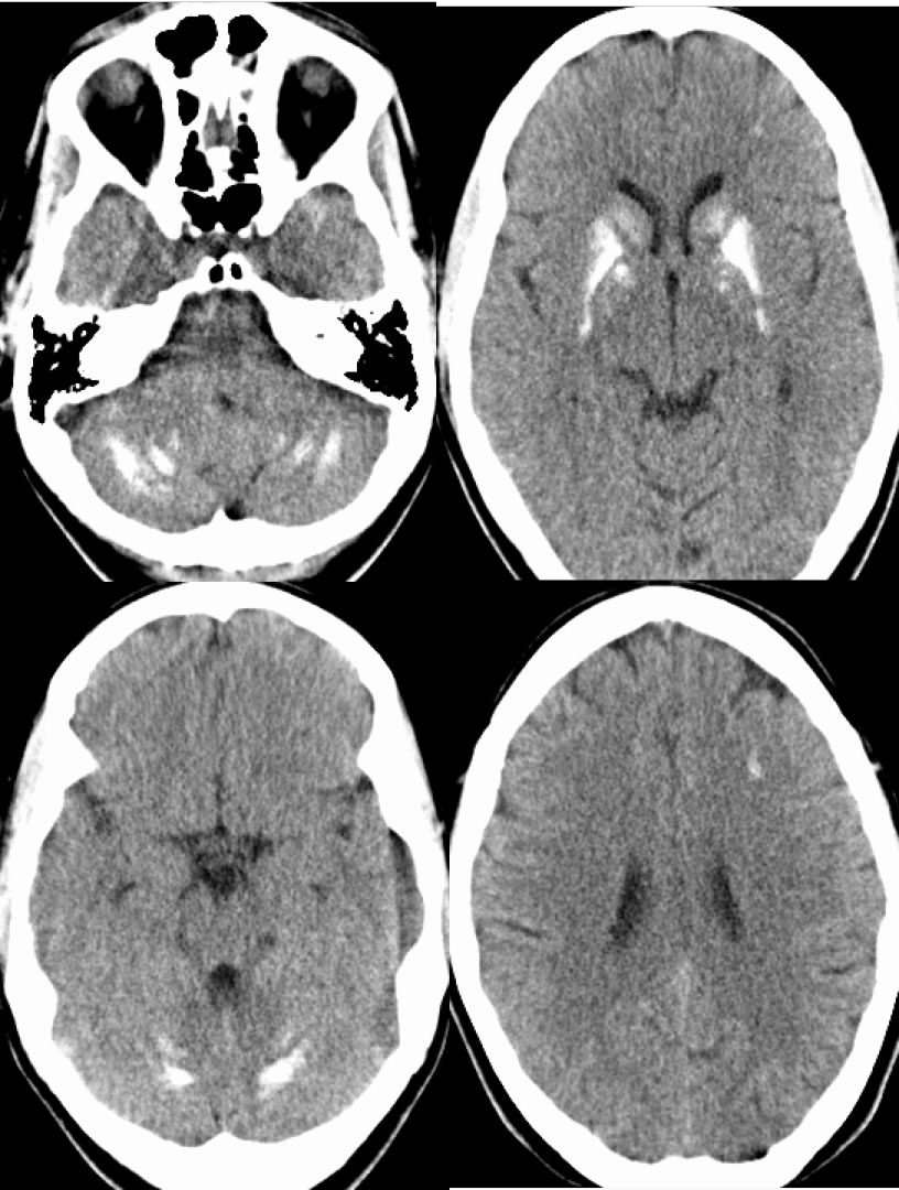
Multiple axial MRI images demonstrate symmetric zones of T1 hyperintensity involving the bilateral caudate nuclei, basal ganglia, and dentate nuclei also extending into the corona radiata. No hemorrhagic staining or diffusion restriction is seen. The corresponding CT examination demonstrates calcification in the regions of MR abnormality, as well as some subcortical calcifications that are not well visualized on MR.
Discussion/Differential Diagnosis:
T1 hyperintensity in the basal ganglia related to liver disease and gadolinium deposition is not typically this extensive and obvious. The symmetry indicates a more diffuse systemic underlying metabolic process, and CT is helpful to demonstrate that this represents calcium rather than other products. Disorders of calcium metabolism including secondary hyperparathyroidism from renal disease should be considered when symmetric parenchymal brain calcifications are seen. Asymmetric calcifications are an entirely separate differnetial diagnostic pathway. Fahr's Disease as a congenital disorder has a similar appearance, but is also thought to be related to abnormal calcium metabolism.
BACK TO
MAIN PAGE