Glomus Jugulotympanicum
Findings:
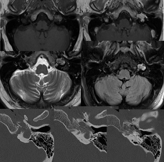
Axial MR images demonstrate an irregular enhancing mass within the region of the left jugular foramen which is slightly hyperintense on T1 weighted imaging, demonstrates a few spotty hypointensities on T2, and demonstrates diffuse enhancement. The CT images demonstrate permeative bone destruction in the region and a lobulated mass component extending into the middle ear cavity.
Discussion/Differential Diagnosis:
The presence of permeative bone destruction with a hypervascular mass in the region of the jugular foramen should raise suspicion for a paraganglioma. The T2 hypointensities are compatible with flow voids substantiating hypervascularity. Schwannoma may occur in the same region, but the poorly defined nature, flow voids, T2 signal, and permeation would be unusual. Other cases are below with additional discussion.
BACK TO
MAIN PAGE