Lumbar Epidural Abscess
Findings:
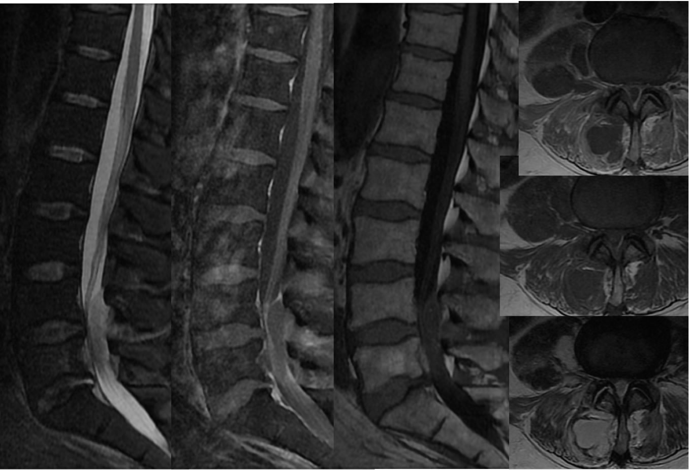
Sagittal T2 fat saturated, sagittal T1 fat saturated post contrast, and sagittal T1 imaging of the lumbar spine demonstrates a rim enhancing complex fluid collection dorsally at the L4-5 level causing severe thecal sac compression. Associated nerve root enhancement is present in the region of thecal sac compression. Axial T1 post contrast, axial T1, and axial T2 weighted images again demonstrate the compressive epidural fluid collection, with additional components of similar complex fluid collection extending within the paraspinal musculature and the right psoas. An enhancing annular fissure is incidentally noted in the left extraforaminal region.
Discussion/Differential Diagnosis:
The differential diagnosis of epidural masses is broad including neoplastic and infectious processes. In this case, a clinical history of IV drug abuse is helpful. The multiloculated mutiseptated appearance, irregular peripheral enhancement, surrounding soft tissue changes, and involvement of paraspinal musculature should strongly raise suspicion for abscess, and discitis/osteomyelitis need not be present. For additional cases and discussion, please refer to the cases below.
BACK TO
MAIN PAGE