Central Neurocytoma
Findings:
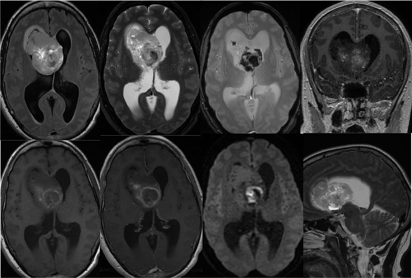
Multiple MR images pre-and post contrast demonstrate a large heterogeneously enhancing mass within the right frontal horn which obstructs the foramina of Monro and causes obstructive hydrocephalus. A zone of heterogeneous diffusion signal correlates with hemorrhagic changes within the mass as seen on the gradient echo sequence. The mass demonstrates mixed zones of hyperintensity and hypointensity on the T1 and T2 weighted sequences.
Discussion/Differential Diagnosis:
The differential diagnosis for this lesion includes central neurocytoma, metastasis, subependymal giant cell astrocytoma, choroid plexus tumor, or meningioma. Subependymoma is less likely due to the size and enhancement. The heterogeneity and hemorrhagic zones would not be expected with meningioma. The attachment to the septum pellucidum, heterogeneity, and location near Foramen of Monro indicates likely central neurocytoma. Additional cases and discussion of central neurocytoma:
-central neurocytoma
-central neurocytoma2
BACK TO
MAIN PAGE