Pituitary Macroadenoma
Findings:
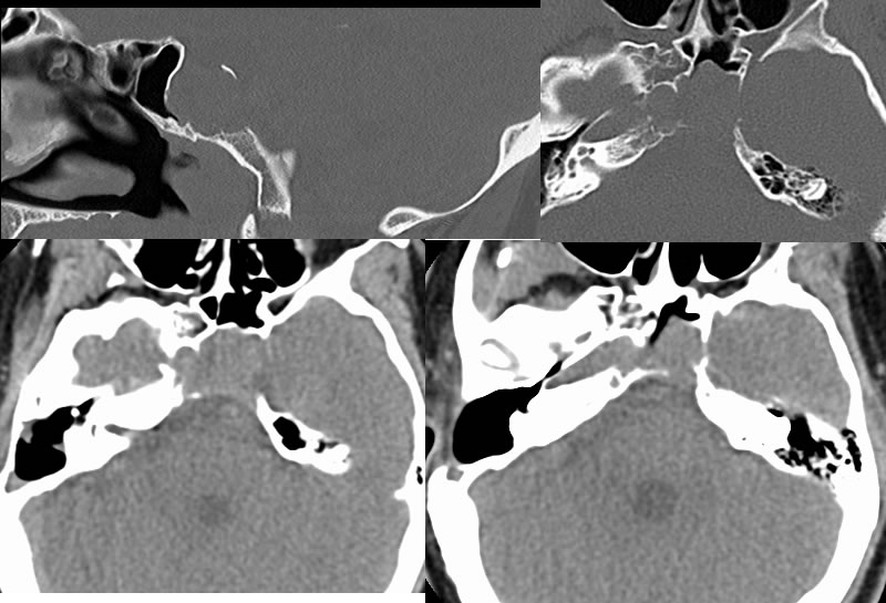
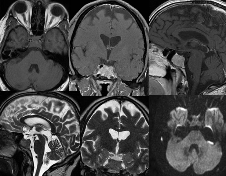
Multiple noncontrast CT images demonstrate a soft tissue mass destroying the central clivus and sella. Some bony remodeling of the sella is seen with mass extending into the sphenoid sinus. The mass is isodense to hyperdense to brain parenchyma on CT. MR imaging demonstrates a heterogeneously enhancing solid mass within the clivus and sella, which demonstrates relative decreased signal on T2 weighted imaging, no definite restricted diffusion, and no significant narrowing of the internal carotids. A thin sclerotic line along the inferior margin of the mass indicates a long-standing process. The pituitary infundibulum is in midline position and there is no suprasellar mass or optic chiasm compression. The mass is inseparable from the bilateral cavernous sinuses.
Discussion/Differential Diagnosis:
The differential diagnosis of a clival soft tissue mass is somewhat broad, with chordoma, chondrosarcoma, and metastatic disease typically considered first. In this case, the signal and attenuation are not typical for chondrosarcoma and chordoma. Smooth bone remodeling and a sclerotic border would not be expected for these three etiologies.
Once a sellar origin is recognized, a pituitary macroadenoma should be considered far more likely than craniopharyngioma. This case illustrates that pituitary adenomas sometimes grow inferiorly and into the cliveus rather than having the more common suprasellar extent. Additional cases and discussion of pituitary adenoma:
-pituitary adenoma 1
-pituitary adenoma 2
BACK TO
MAIN PAGE