Basilar Artery Thrombosis
Findings:
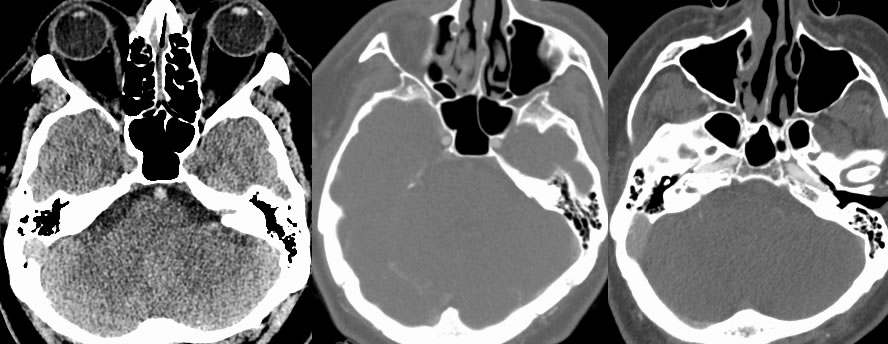
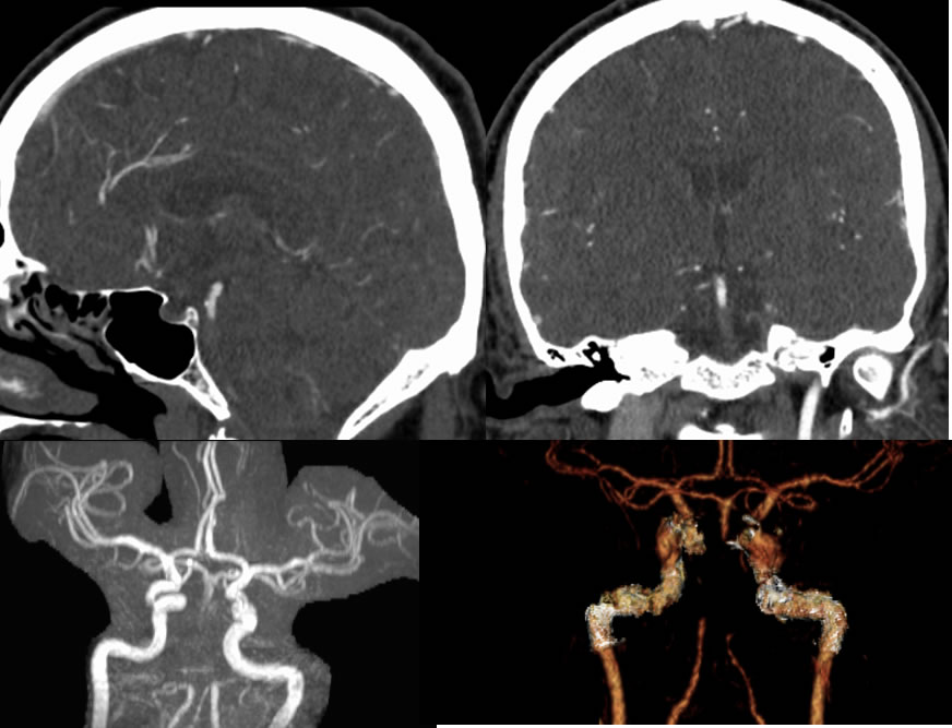
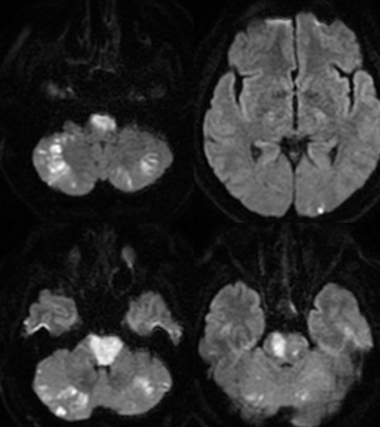
Axial noncontrast CT image demonstrates asymmetric hyperdensity of the basilar artery compared to the parasellar internal carotids. Multiple CTA images including reformats demonstrate occlusive thrombosis of the basilar artery. Diffusion weighted imaging demonstrates mutiple zones of acute infarction involving the bilateral cerebellum, pons, and left occipital lobe in posterior circulation distributions.
Discussion/Differential Diagnosis:
Asyemmetric CT hyperdensity of an intracranial vessel should raise concern for thrombosis, taking care to account for artifactual changes in density that may occur adjacent to bone interfaces. In this case, there is clear asymmetric hyperdensity of the basilar artery compared to other arterial structures on this image. The CTA makes the occlusive thrombus more obvious. Additional similar cases and discussion are below:
-Basilar artery thrombosis
-Basilar artery thrombosis 2
BACK TO
MAIN PAGE