Neuromyelitis Optica
Findings:
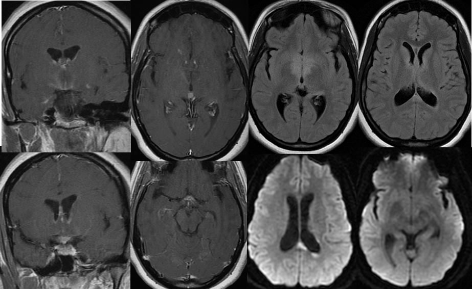
Multiple MR images demonstrate patchy nearly symmetric signal abnormalities involving the bilateral hypothalamus, periventricular regions, optic chiasm, and corpus callosum splenium. The corpus callosum splenium lesions demonstrate restricted diffusion. There is patchy enhancement of this process which is predominantly peripheral, and extensively involves the optic chiasm as well as the subependymal margins of the frontal horns and lateral margins of the hypothalamus. No significant mass effect is seen.
Discussion/Differential Diagnosis:
The differential diagnosis for this process is somewhat narrow, including a demyelinating type process and metabolic processes. The central midline involvement including the optic chiasm is thought to be fairly characteristic for neuromyelitis optica. Other cases and discussion of neuromyelitis optica are found below:
-neuromyelitis optica
-neuromyelitis optica 2
BACK TO
MAIN PAGE