Diffuse Axonal Injury (DAI)
Findings:
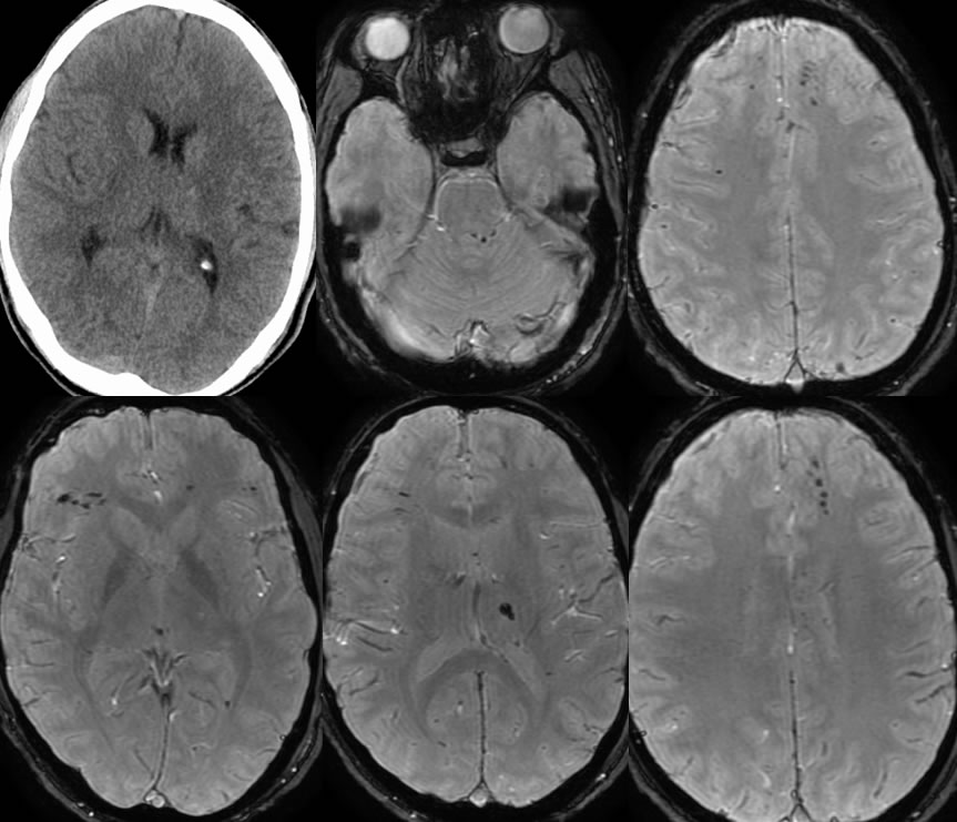
On CT, there is a subtle punctate hyperdensity in the left thalamus near the genu of the internal capsule. The susceptibility weighted MRI imaging demonstrates innumerable punctate foci of hemorrhage within the bilateral frontal lobes, left thalamus, left corpus callosum splenium, right occipital lobe, in a characteristic pattern and distribution for multifocal hemorrhagic shear injuries. The focus of hemorrhage within the superior left thalamus correlates with the zone of hyperdensity seen on CT. There is trace hemorrhage within the occipital horn of the left lateral ventricle. There is minimal subtle shear injury within the left superior cerebellar peduncle.
Discussion:
This case illustrates the importance of MR for demonstrating DAI, also known as shear injury, which may be significantly underestimated on CT. DAI should be considered in any head trauma patient with less than expected mental status and recovery for head CT appearance. Discussion of DAI is found on other cases on this site:
This case was prepared with the assistance of Joshua Hall, UC undergraduate
BACK TO
MAIN PAGE